Alpha Prolipsis' Medical Services
Your reports will be available within 24 hours of receiving the sample
Over the past few months, artificial intelligence (AI) has been seemingly everywhere in the news. Some researchers have portrayed the increasing capabilities of AI algorithms as an exciting boon to research, clinical decision-making, and medical education. Other experts have become so alarmed by some of its applications, such as image generators and chatbots that mimic human conversation, that they have urged a 6-month pause on training powerful AI systems to better assess the risks. Some ethicists, in turn, have pointed to more immediate concerns, such as reinforced biases already being baked into the technology.
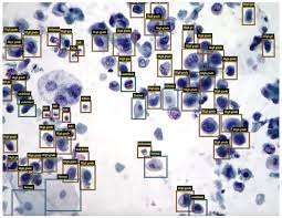
In visual fields such as cytopathology, current AI applications are mostly geared toward identifying patterns and other data in pathology samples, such as Papanicolaou tests. Three cytopathologists who spoke to CytoSource say that they see exciting possibilities with the technology but caution that multiple challenges in optimizing data, minimizing bias, and redefining clinical roles must be resolved before it can reach its full potential.
Brie Kezlarian, MD, an assistant attending pathologist at Memorial Sloan Kettering Cancer Center in New York City,says that using AI as an adjunct to help cytologists to identify cells makes a lot of sense. Gynecologic applications have been particularly useful because of the narrow diagnostic range and the task of identifying fairly rare abnormal cells against a background of normal ones, she says. AI could prove similarly adept at classifying cells or conducting rapid onsite evaluations with other relatively homogeneous cytology preparations, she notes.
“In my opinion, the best mechanism going forward is a marriage of the benefit you get from an automated system and the benefit you get from a human,” Dr Kezlarian says. Under that scenario, AI would flag cells of interest, and then a cytologist would conduct quality control in determining whether the AI-rendered classifications are indeed correct. “I think that it’s important to know what AI can do and what AI can’t do, and that ultimately I’m responsible for the case regardless of whether AI touched it or not,” she says.
One recent review noted that, although most AI efforts have focused on histopathology images, commercialized models have been used extensively for automated Papanicolaou test screening.1 Efforts have expanded on whole slide images in both gynecologic and nongynecologic cytopathology as well. A 2022 article by researchers in Korea reviewed many of the latter applications, including thyroid, bladder, lung, breast, ovarian, pancreatic, prostate, and pleural effusion cancers. Most of the studies focused on classification and segmentation tasks, the authors found. They concluded that “although most of the studies showed impressive results, the sizes of the training and validation datasets were limited.”2
Review coauthor Yosep Chong, MD, associate professor of pathology at the Catholic University of Korea in Seoul, says that the utility of cytology is often seen by other specialists as a good screening tool, but one with limited information and diagnostic accuracy. In Korea, however, cytopathologic examinations are growing at a rate of approximately 7%–10% annually; this is driven by the country’s aging population and its increased risk of cancer. “Cytologic exams are cheap, rapid, less invasive, easy to collect, and easily repeatable,” Dr Chong says. “With the aid of AI, the diagnostic performance of cytologic exams can be dramatically enhanced, potentially replacing histologic confi rmation by biopsy or excision, as well as additional tests, and even molecular tests. This could ultimately reduce medical costs.”
Liron Pantanowitz, MD, chair of pathology at the University of Pittsburgh Medical Center in Pennsylvania and president of the American Society of Cytopathology, says that AI’s capabilities in the field have steadily increased over the past 5 years. Under the technology’s broad umbrella, algorithms can vary widely in the human input that is required. One deep learning algorithm known as a convolutional neural network can analyze patterns with little or no human involvement. Relying on what is known as “unsupervised learning,” however, requires a much larger data set.
Some experts have suggested that a minimum of 10,000 images may be necessary to build robust and accurate data sets for deep learning applications. One way to overcome the limitations, Dr Pantanowitz says, would be to include periodic supervised learning sessions. Separately, augmented techniques could reuse sections of an image or rotate them by 90° to create new images for training sets.
The algorithms, of course, are only as good as the input; “garbage in, garbage out” is a common mantra in the field. As AI progresses, both Dr Kezlarian and Dr Pantanowitz say that researchers will need to focus on standardizing cytology preparations from laboratory to laboratory; on obtaining fast, reliable, and highquality digital images; and on ensuring that the data sets used to train the algorithms are as representative and bias-free as possible. “At the end of the day, we’re the specialists. We know what we need to make diagnoses,” Dr Kezlarian says. “So, it only makes sense that cytopathologists are fundamentally involved at all levels of the development and deployment of AI in cytopathology.”
One unresolved challenge is how to design robust quality control and quality analysis for AI algorithms when it is not always clear what the “black box”
“With the aid of AI, the diagnostic performance of cytologic exams can be dramatically enhanced, potentially replacing histologic confi rmation by biopsy or excision, as well as additional tests, and even molecular tests.” Yosep Chong, MD
formulas are focusing on to classify cellular images. The fi eld also needs more data on outcomes, Dr Pantanowitz says, to help to answer whether AI applications are indeed helping with cytology workloads and patient care.
At least in the near future, Dr Chong, Dr Kezlarian, and Dr Pantanowitz agree that AI is not likely to replace humans in cytology laboratories. “Even with the increasing use of AI, there will always be problems to solve and responsibilities to take that only human experts can handle,” Dr Chong says. Because of the surging demand for cytopathology, he adds, AI applications may help to address increasing workloads and staffi ng shortages.
Dr Kezlarian says that she hopes that AI in medical settings will actually give humans more time to do the things they are good at, such as critical thinking, taking a holistic view, and creative problemsolving. “A human has the ability to think outside the box,” she notes, whereas an algorithm can do only what it has been taught. The role of cytologists, meanwhile, is expanding to include more molecular testing and investigation of patient histories, and algorithms that take on the burden of classifying cytology images could help to relieve some of the pressure. “Certainly, if AI can make our work better, faster, and more effi cient, then we have an obligation to do so, I think,” she says.
Dr Pantanowitz agrees. “We have always been leaders in adopting new technology,” he says of cytopathology. AI technology, he adds, could further empower the cytology workforce in exciting ways. “It really untethers the cytologists from the microscope,” he says. That means they are not necessarily tied to a single location. If they can log on digitally to screen slides, they can redistribute the workload and extend their reach.For the past 5 years, in fact, Dr Pantanowitz says, AI has become his “microscope” for exploring new research questions.
“There’s much more than meets the eye in an image,” he says, and AI is helping to reveal that additional information and open new fi elds such as spatial biology applied to cancer research.
If it proves trustworthy, AI technology will undoubtedly allow for more rapid screening. Dr Pantanowitz says that shift, in turn, will require the fi eld to set new workload limits. To help to make recommendations and address ethical issues, he has assembled a digital pathology and AI task group for the American Society of Cytopathology. The group’s white paper will be presented at the society’s conference in November.
Dr Chong, for his part, has led an effort to develop AI models from thousands of whole slide images of respiratory tract samples, pleural fl uid, ascites, urine, and fine-needle aspirations from 200 registered pathology laboratories across Korea. Eventually, he believes that AI could serve as a quality control tool to help to ensure patient safety and “help predict histologic subtypes, related mutations, treatment responses, and survival rates without additional tests.”
In the meantime, Dr Chong says, pathologists may be adopting a more practical view of the technology. At the 2023 United States and Canadian Academy of Pathology conference, he noticed a signifi cant shift: “Most doctors now view AI as an assisting tool rather than a total problem solver.” Like Dr Kezlarian, he says they understand that they still need to verify AI-generated results and assume full responsibility for the final diagnosis.