Alpha Prolipsis' Medical Services
Your reports will be available within 24 hours of receiving the sample
ALPHA PROLIPSIS
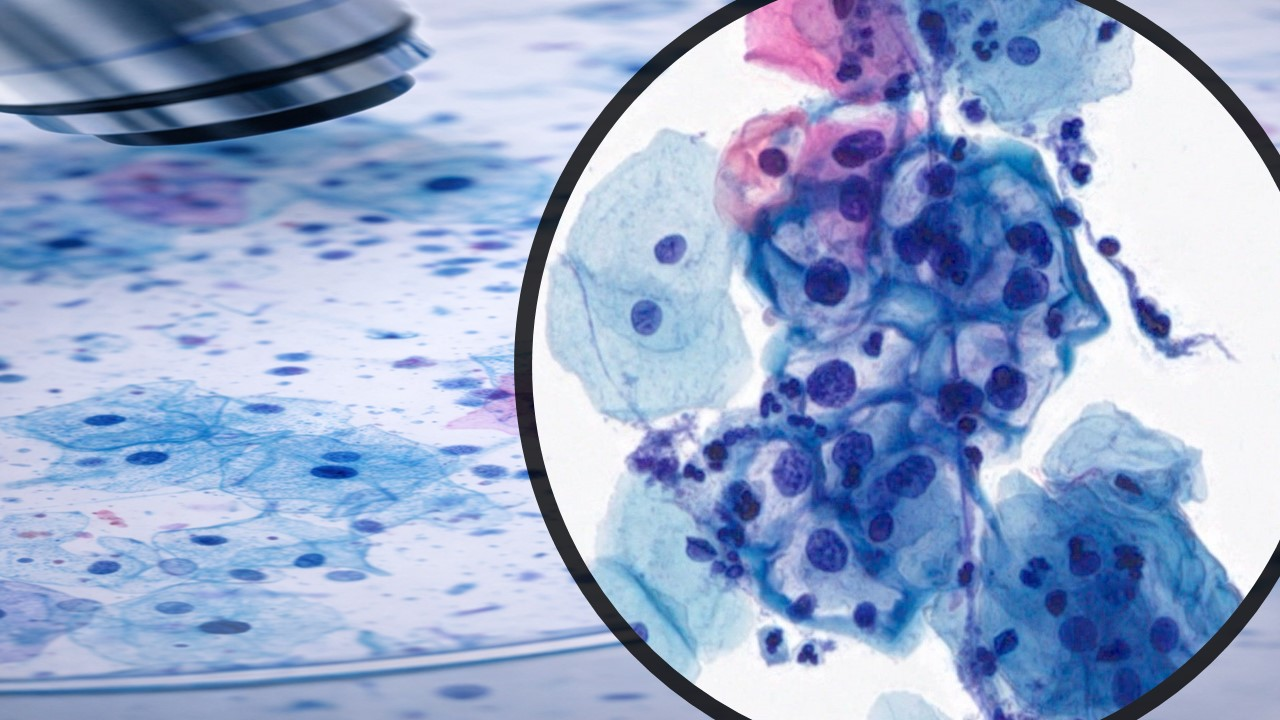
ANAL PAP SMEAR
Invasive squamous cell cancers of the anal canal are associated with certain types of human papillomavirus (HPV) infection, most notably, HPV 16 and HPV 18. Although the overall incidence of anal cancer in the general population of the United States is low (approximately 0.8/100,000), its incidence varies considerably depending on the presence of risk factors such as smoking, multiple sexual partners, HPV infection, and receptive anal intercourse. HIV infection confers an additional risk for development of anal cancer.
Analogous to cervical tissue, the anal epithelium at the dentate line has a transformation zone between squamous and columnar epithelia; this transition zone is subject to infection with and neoplastic transformation by HPV. Precancerous lesions of the anal squamous epithelium can develop and are classified as low- or high-grade according to identical Bethesda criteria and nomenclature developed for grading cervical lesions.
RISK OF ANAL CANCER
HIV infection is an independent risk factor for anal neoplasia. Individuals with a history of long cumulative periods of immunosuppression (CD4 count <200 cells/mm3) and/or high viral replication may be at higher risk for developing anal cancer. Behaviors that have been associated with anal neoplasia include multiple sexual partners, smoking, and unprotected vaginal or anal intercourse.
A. HIV-Infected Women
The risk of anal cancer in women regardless of HIV status is highest among those who have had 10 or more sexual partners, those with a history of anal or genital warts, gonorrhea, or cervical neoplasia, and those whose sexual partners have a history of a sexually transmitted infection. A history of receptive anal intercourse before 30 years of age or with multiple partners increases the risk of anal cancer.
Women with HIV infection are significantly more likely to have abnormal anal cytology/histology (31%) compared with non-HIV-infected women (9%). Data from the United States AIDS-Cancer Registry Match calculate an increased relative risk of 134 for anal cancer among HIV-infected women younger than 30 years of age, and a relative risk of 12 for women aged 30 to 39 years compared with aged-matched non-HIV-infected women. From 1973 to 1989, the incidence of anal carcinoma increased by 35% and has been increasing at a rate of 2% per year. In one report, 26% of HIV-infected women had abnormal cervical cytology, and, of those women, 44% also had abnormal anal cytology.In the ongoing Study to Understand the Natural History of HIV/AIDS (SUN), the prevalence of HPV in the cervix and anus was 86% and 93%, respectively, and for HPV types that carry a high risk for malignancy, the prevalence rates were 68% and 85%, respectively.A history of anal sex was not predictive of an abnormal anal cytology. These results, although not completely independent of a history of anal intercourse, are explained by the anatomical proximity of the anus and the genital tract. HPV exposure of either anatomical site can result in tracking and infection of the other site.
B. HIV-Infected Men
Prior to the onset of the AIDS epidemic, the risk of anal cancer was estimated to be as high as 35/100,000 among men with a history of receptive anal intercourse. The risk in HIV-infected men who have sex with men (MSM) is between 70/100,000 and 144/100,000,13,14 and this risk does not appear to decrease with effective immune reconstitution as a result of HAART.
Among men who do not engage in receptive anal intercourse, the risk of anal cancer is associated with 10 or more sexual partners and a history of anal warts, syphilis, or hepatitis.7 In MSM, receptive anal intercourse is the most common risk factor and compounds the risks noted above.
II. SCREENING AND DIAGNOSIS
Recommendations:
At baseline and as part of the annual physical examination for all HIV-infected adults, regardless of age, clinicians should:
Clinicians should refer women with cervical HSIL and any patient with abnormal anal physical findings, such as warts, hypopigmented or hyperpigmented plaques/lesions, lesions that bleed, or any other lesions of uncertain etiology, for high-resolution anoscopy and/or examination with biopsy of abnormal tissue.
Clinicians should obtain anal cytology at baseline and annually in the following HIV-infected populations:
Delayed diagnosis of anal cancer is common. MSM, the group with the highest risk of anal cancer, often have benign conditions such as fissures or infections that may mask the diagnosis. Rectal bleeding, which is the most common presenting symptom of anal cancer, is often attributed to hemorrhoids. Only 30% of patients have pain or the sensation of an anal mass. As with cervical carcinoma, the precancerous stages of anal intraepithelial neoplasia are generally asymptomatic until there is invasion beyond the epithelial basement membrane, concurrent with tumor enlargement. Patients may complain of thickening and irritation of perianal skin, itching, bleeding, tenesmus, pain with defecation, constipation, change in stool caliber, or receptive anal dyspareunia. Upon visual inspection, clinicians should examine for abnormal anal physical findings, such as warts, hypopigmented or hyperpigmented plaques/lesions, or lesions that bleed.
Anal Cytologic Screening
1. Rationale
The risk of anal cancer in the MSM population (between 70/100,000 and 144/100,000) is double the rate that prompted the recommendations for universal cervical screening of women. Universal cervical Pap screening for adult females was recommended in the United States in the 1960s before either the pathophysiology or oncogenesis of HPV was elucidated. There has never been a randomized control trial for standard cervical Pap screening, nor would it be ethical to conduct such a study today. The efficacy of Pap screening in preventing cervical malignancies rests exclusively on decades of epidemiological data for validation. Given that 1) it is unlikely that a 10-year clinical trial demonstrating the efficacy of obtaining anal cytology in any HIV-infected population will ever be performed and 2) HPV-initiated carcinomas are clearly preventable malignancies, this committee recommends obtaining routine anal cytology in populations that are clearly at risk.
Anal Pap testing using a Dacron swab is a well-validated technique with comparable sensitivity and specificity to cervical cytology. Pap test screening of the cervix has led not only to a markedly decreased incidence of invasive cervical cancer but also to an understanding of how precursor lesions of the cervical epithelium can progress to invasive disease. As with cervical cancer screening, cytologic screening of the anal canal is expected to reduce the incidence of invasive anal cancer and allow the detection of precancerous dysplastic lesions or treatable early invasive disease. Pap tests of the anal canal are simple to perform, clinically effective, and cost-effective to reduce the incidence of invasive disease in high-risk individuals.
2. Technique
There is no preparation necessary before obtaining anal cytology. If the digital rectal examination is performed in conjunction with anal cytology and/or HRA, the cytology must be obtained first, before lubrication is introduced into the anal canal. Patients should not have received an enema or engaged in receptive anal sex within 24 hours before sampling because these activities can adversely affect specimen quality.
The standard technique used in obtaining anal cytology is as follows: a Dacron swab (a cotton swab will not yield accurate results) is moistened with sterile or non-sterile water. The anus is spread with the index and thumb of the non-dominant hand so that the anoderm pouts out. The swab is then gently inserted into the anal canal as far as it will go, until it hits the wall of the rectum. If the swab does not go in easily, the angle of insertion should be adjusted. The presence of external hemorrhoids may cause resistance; in this case, different insertion points should be tried until the anal canal is easily accessed. The swab must be inserted above the squamocolumnar transition zone, which is approximately 2 cm (1 inch) from the anal verge.
The swab is then slowly moved in and out without completely withdrawing it, while rotating it in a spiral motion and applying mild pressure to the anal wall. After several rotations, the swab should be withdrawn and immediately immersed in methanol-based preservative-transport solution. Feces or traces of blood on the swab will not affect the result. The swab should be agitated in the solution for 60 seconds to transfer cells from the swab to the medium.
Slides made by the thin-layer liquid-based cytology process display a thin, uniform layer of cells at a controlled density. Red blood cells and mucus are removed while the background pattern and the cell clusters are preserved. The use of this process results in increased detection of abnormal cytology. The absence of columnar cells in the sample does not affect the validity. The sensitivity, specificity, and predictive value do not hinge on the presence or absence of these columnar cells; cytological specimens should not be rejected solely on this basis. If only anucleated squamous cells are present on the sample, the swab was not inserted far enough into the anal canal, and the specimen is inaccurate or non-diagnostic.
3. Follow-up
Recommendation:
Clinicians should refer patients with abnormal anal cytology for high-resolution anoscopy and/or examination with biopsy of abnormal tissue.
}Anal cytology has a high sensitivity (95%) for detection of dysplasia (ASIL) but a low specificity (50%) for predicting the severity of the abnormality in subsequent biopsy. Even patients with cytologic diagnoses of ASC-US and LSIL have a significant risk (46% to 56%) of being diagnosed with HSIL at biopsy. Although the appropriate follow-up for abnormal anal cytology remains an active area of investigation.
Health Tips
The ABCs of anal-rectal cytology
The use of anal-rectal cytology is becoming more common for evaluating human papillomavirus-related disease of the anal canal, especially in at-risk populations—principally men who have sex with men and those with HIV disease. However, few laboratorians are familiar with anal-rectal cytology, or ARC. This article is a primer on ARC.
Squamous cell carcinoma, or SCC, of the anus has become a serious health issue in the gay community in recent years. Because advances in combination drug therapy have increased life expectancy in those with HIV disease, neoplasms that develop slowly over time, such as SCC, have had the chance to persist and progress to cancer. Risk factors for developing anal SCC, in both men and women, include anogenital human papillomavirus infection, anal receptive intercourse, multiple sexual partners, history of sexually transmitted disease, and history of anal condyloma. Women with anal SCC are likely to have a prior history of cervical intraepithelial neoplasia or cervical carcinoma. Other causes of immunosuppression, including steroid therapy and renal transplantation, are also associated with an increased risk of all types of anogenital carcinoma.
The incidence of anal SCC in men who have sex with men is currently estimated to be 35 per 100,000.1 Interestingly, this is approximately the same incidence as that of SCC of the uterine cervix before the institution of screening programs using Pap smears. Routine Pap smear screening has reduced the rate of cervical cancer from 40 per 100,000 to eight per 100,000. Now, in the AIDS era, the anal cancer incidence in HIV-positive men who have sex with men is estimated to be double that and may be as high as 70 per 100,000.2 Carcinoma of the anus now represents about 1.5 percent of cancers of the digestive tract. The American Cancer Society projects that about 4,010 new cases will be diagnosed in the United States in 2004, up from 3,400 cases in 2000, and that about 580 will die of the disease this year.
The etiology and pathogenesis of anal squamous neoplasia are analogous to those of squamous neoplasia of the uterine cervix. Anal squamous intraepithelial lesions, or ASIL, arise in the transition zone of the anus, an area extending from the squamous mucosa of the anus through the dentate line to the squamo-columnar junction with the rectal columnar mucosa. In this area of transition, there is active changeover of columnar epithelium to squamous epithelium through the process of squamous metaplasia. This process is accelerated by trauma, healing, and repair such as might be expected to occur with receptive anal intercourse.
The transition zone is also peculiarly susceptible to infection with human papillomavirus, or HPV, particularly by the genital HPV types associated with benign, low-risk genital types leading to condyloma, and the intermediate/high-risk genital types associated with ASIL and SCC. Anal condylomas are most often associated with HPV types 6/11, while HPV type 16 is the most common HPV-type in ASIL and SCC. These are the same viral associations that are noted in the uterine cervix. In HIV-infected patients, immunosuppression acts to increase susceptibility to infection and increases the risk that HPV infection will persist and that the associated lesions will progress. Approximately 95 percent of gay men with HIV, and 65 percent of gay men who are HIV-negative, can be shown to be infected, at some time, with HPV in the anal canal or in the peri-anal skin.3 In HIV-positive men, the highest risk is determined by low baseline CD4 counts and evidence of persistent HPV infection with intermediate or high-risk HPV types, and infection with multiple HPV types.
Because of the effectiveness of cervical cancer screening in women, a number of groups around the country have been investigating the feasibility and effectiveness of screening the anal-rectal transition zone for anal SCC and its precursors, in much the same way that we now screen the uterine cervix. A number of studies have been completed, and it has been demonstrated that anal-rectal cytology is an accurate and cost-effective screening method for detecting ASIL.4-6 In one of the largest prospective studies, by Palefsky et al, 346 HIV-seropositive and 262 HIV-seronegative men who have sex with men were studied and correlated using cytology, HPV testing, anoscopy, and biopsy.3,7,8Abnormal cytology was defined as ASCUS (atypical squamous cells of undetermined significance) and above. The sensitivity of cytology for detecting biopsy-proven ASIL was 69 percent in HIV-positive men and 47 percent in HIV-negative men at the initial visit. Repeat cytology increased sensitivity to 81 percent and 50 percent in these respective groups of men.6 These rates are comparable to those seen in screening of women for cervical cancer. Accumulating evidence also suggests that all people with HIV disease, regardless of their gender and sexual orientation, have high rates of HPV-related anogenital disease and may benefit from anal cytologic screening.9,10
Collection and preparation of ARC Taking an anal-rectal sample for cytology is a simple procedure. It does not require the use of an anoscope. No special preparation is needed for the patient, though the patient may be advised to refrain from receptive anal intercourse or the use of intra-anal preparations before examination.
An ARC sample can be collected with the patient in either the lateral recumbent or dorsal lithotomy position. If the patient is already being seen for a gynecologic exam, lithotomy is often more convenient; the ARC sample can be collected before or after the gynecologic exam. For male patients or if a gynecologic table with stirrups is not available, lateral recumbency is more commonly used. Here the patient is lying on his side, with his knees drawn up toward his chest.
To collect an ARC sample, a tap water-moistened Dacron swab is used. The Dacron swab is inserted about 5-6 cm into the anal canal past the anal verge, into the rectal vault. This is done without direct visualization of the anal canal. Firm lateral pressure is applied to the swab handle as it is rotated and slowly withdrawn from the anal canal, inscribing a cone-shaped arc. Care should be taken to ensure that the transition zone is sampled. A swab or smear of the peri-anal skin is an unsatisfactory sample for ARC. Some have reported using a cytobrush for collecting ARC, but the cytobrush may be more uncomfortable for the patient. Avoid using cotton swabs on a wooden stick because the handle may break and splinter during collection.
For liquid-based cytology, the swab is then placed in the preservative vial and agitated vigorously several times to release the cellular harvest. If liquid-based cytology is not available, the swab can be smeared onto a glass slide and then spray-fixed as per the procedure for conventional cervical Pap smears.
In the laboratory, there are a variety of ways that liquid-based samples can be prepared. For ThinPrep ARC, the samples can be collected in CytoLyt and processed using the blue filter; they are run on sequence No. 1 for superficial cells or sequence No. 3 for fluids/FNA. Another "off-label" alternative for ThinPrep ARC, preferred by one of the authors (TD), is to process these samples similarly to cervical samples. The sample is collected in PreservCyt, which can be used for brushings from any body site (James Linder, MD, Cytyc Corp., personal communication), and then processed using the white filter and the gyn program (sequence No. 4 for gyns) on the ThinPrep processor. The larger pore size of the white filter may help to reduce the amount of fecal contamination occasionally encountered with ARC. ThinPrep specimens are displayed in a 20 mm diameter circle.
For Tripath specimens processed using the PrepStain System, the sample is submitted in CytoRich Red Fixative and processed using standard nongyn sample procedures (Karen Atkison, CT, TriPath Imaging Inc., personal communication). This results in a preparation with a diameter of 13 mm. Alternatively, after collection in CytoRich Red, the sample is vortexed for five minutes, then cytocentrifuged using a Hettich Cytocentrifuge bucket which results in a 16 mm diameter circle (Paul Elgert, CT, New York University, personal communication).
Billing for ARC ARC is billed using CPT codes for nongynecologic samples (88108 for liquid-based cytology and 88104 for conventional brushings and 88160 for anal-rectal smears using a Dacron swab). A new CPT code (88112—selective cellular enhancement technique with interpretation) for nongyn liquid-based preparations was instituted this spring.
Currently, ARC cannot be billed as a screening test, as are cervical Paps. There is no CPT code for this category of sample as a screening test. Third-party payers will reject a bill for ARC if CPT codes for gynecologic cytology are used, especially if from a male patient.
Interpretation and reporting ARC is interpreted and reported using the Bethesda 2001 guidelines, modified for this site.11 Both ARC and cervical cytology are used to evaluate cytologic changes associated with HPV-related precursor lesions and cancers; similar cytomorphologic criteria are used. Learning to evaluate an ARC is akin to learning a new dialect, rather than a whole new language. Different nuances are seen with ARC, but those who evaluate cervical samples should find ARC especially interesting because the target populations have such high rates of abnormalities.
One of the challenges of ARC is the collection of an adequate sample. Procedures for collecting these samples have been noted here. Because the anal canal is not directly visualized at the time of cytology, the clinician collects these samples "blindly." Feedback from the laboratory can be helpful in guiding the clinician to collect an adequate sample. Although there is little published data on what constitutes an adequate ARC, those who look at these samples frequently have developed criteria. The cellularity of most ARC, collected by experienced clinicians, equals that of cervical samples. ARC typically consists of anucleated squames from the keratinized portion of the canal, nucleated squamous cells, squamous metaplastic cells, and rectal columnar cells. In general, liquid-based cytology is preferred over conventional smears for evaluating ARC because there is increased cellular yield and improved cell preservation. Liquid-based preparations also reduce fecal material and bacteria that can obscure cellular detail. The air-drying and mechanical artifacts that are commonly encountered with conventional anal smears are eliminated with liquid-based cytology.
}The recently published second edition of the Bethesda System atlas contains guidelines for specimen adequacy for ARC.11 As a guide, the minimum cellularity for an adequate ARC sample is approximately 2,000 to 3,000 nucleated squamous cells. This is equivalent to one or two nucleated squamous cells per high-power field (hpf) for ThinPreps (with a diameter of 20 mm) and three to six nucleated squamous cells/hpf for TriPath's PrepStain System (with a diameter of 13 mm) using a standard 40? objective. Samples with scant cellularity or those that consist primarily of anucleated squames are unsatisfactory for evaluation. The presence of squamous metaplastic and rectal columnar cells is reported as an indication that the anal transition (transformation) zone has been sampled.
The criteria used to interpret HPV-related lesions of the anus and cervix are essentially the same. However, cellular degeneration and keratinization of the squamous intraepithelial lesions are more frequently encountered on ARC than on cervical Paps.
Low-grade SIL, or LSIL, is characterized by the presence of koilocytes and by superficial and high intermediate cells with an increased nucleus to cytoplasmic (N/C) ratio (See Fig. 1). Bi-nucleation is frequently seen. The nuclear membrane is often angulated and irregular. Chromatin clumping and hyperchromasia may be seen. Atypical parakeratotic cells may be numerous. Degenerated koilocytes consisting of an "empty halo" are frequently encountered.
In high-grade SIL, or HSIL, the abnormal squamous cells are of low intermediate or immature squamous metaplastic type (See Fig. 2). High-grade cells have high N/C ratios with nuclear enlargement, albeit minimal at times. Nuclei typically have coarse chromatin and wrinkling or irregularity of the nuclear membrane. Chromatin margination and clearing may also be noted in some nuclei. Nucleoli are inconspicuous. The high-grade cells may be numerous and are usually present as single, discohesive cells or as small clusters. Sheets and synctitia are not a common feature of HSIL in ARC. A mixture of characteristic HSIL with atypical surface parakeratotic cells is also common, representing a keratinizing HSIL. Invasive squamous cell carcinoma is difficult to diagnose prospectively, in our combined experience, and most cases are classified as HSIL. Diathesis is not present. The lack of tumor diathesis and findings specific to invasion may be related to the fact that the rectum is not a closed system and that exfoliated material and cellular debris may be excreted with feces. Cytologic findings in advanced SCC have not been well studied.
Reactive and reparative changes are relatively uncommon in ARC, except in cases associated with inflammation and ulceration, such as seen with herpes virus infection. Cells with enlarged nuclei that lack chromatin abnormalities sufficient for an interpretation of SIL should be classified as ASC-US or ASC-H depending on the cell size and N/C ratio. Samples with only rare atypical squamous cells, but lacking definitive cellular evidence for SIL, should also be interpreted as ASC-US.
In our experience, glandular lesions of the anal canal diagnosed by cytology are very rare in the patient populations targeted for screening. One case of atypical glandular cells on ARC was found to come from a hyperplastic polyp of the rectum on further evaluation of the patient. Theoretically, adenocarcinomas of the lower rectum may be sampled with ARC. These could be expected to occur in an 'older' age group but will probably be encountered as the target populations for anal screening age.
Coexisting infections may also be noted in ARC, particularly in the HIV-positive patient. These include herpes virus and cytomegalovirus infections, and Candida. Other organisms, such as amebic cysts and trophozoites, pin worm eggs, and strongyloides, have been encountered.
Triage and treatment It is recommended that all patients with ASC-US or higher on ARC be triaged to anal colposcopy, also known as high-resolution anoscopy, or HRA.12 The role of HPV testing in the triage of ASC-US or higher on ARC has not been well studied. Moreover, the hybrid capture technology used for cervical samples has not been FDA-approved for use in ARC. It should be noted that the rates of HPV positivity are quite high in the populations targeted for screening using ARC. However, HPV testing may prove useful in triage, because of its good negative predictive value, and in the followup of patients with treated anal disease (Barbara Winkler, MD, Stephen Goldstone, MD, and Elaine Alt, MD, unpublished data).
High-resolution anoscopy is the equivalent of cervical colposcopy.13 Since the anus tends to be more sensitive to vinegar than the cervix and vagina, a three percent acetic acid solution is used (as compared with the standard five percent solution used for cervical colposcopy) along with a disposable anoscope. After the anoscope is inserted into the anal canal, a 4-in. by 4-in. piece of gauze sponge, soaked in the three percent vinegar solution, is wrapped around the handle of a Q-tip or scopette and introduced into the anal canal via the anoscope. Once the swab is in place, the anoscope is removed, and the vinegar solution is allowed to saturate the epithelium for a couple of minutes. The swab is then removed, the anoscope reinserted, and the colposcope focused for magnified visual inspection.
During the anoscopic evaluation, the clinician looks for the classic colposcopic changes of acetowhite epithelium, or AWE, and the vascular changes of punctation and mosaicism. Areas of AWE tend to be well demarcated. Many lesions, particularly those of low grade, have architectural contour changes and present as cauliflower-like, papillary, or macular/ papular areas of AWE. Vascular changes are also observed, particularly punctation and mosaicism, and are more commonly associated with HSIL. In general, high-grade lesions tend to be flat, though they may have a warty or exophytic appearance. High-grade SIL may also be identified in the deeper portions of more typical condyloma and LSIL.14 Anoscopic evaluation of the keratinized portion of the anal canal and the surrounding perianal skin should also be included. On keratinized epithelium, the anoscopic changes associated with SIL are similar to those seen on vulvar colposcopy. A digital examination of the anal canal should be included as an essential part of the complete evaluation.
Just as with high-grade cervical lesions, high-grade ASIL is treated to prevent the development of invasive carcinoma. Ablative and excisional techniques, similar to those used for the cervix, can be used.12 For invasive cancers, early detection of anal carcinoma is essential because tumor size is such an important prognostic factor.15 Tumors less than 2 cm are curable with local therapy in 70 to 90 percent of cases. The cure rate drops to 50 percent for tumors with nodal involvement or tumors greater than 5 cm.
Conclusions There is ample evidence in the literature to support the use of ARC as both a screening and diagnostic tool for evaluating patients who are at risk for developing HPV-related lesions of the anal canal. The approach to caring for these patients is multidisciplinary and involves clinicians from many different disciplines of medicine, including family medicine, internal medicine, infectious disease, dermatology, gynecology, surgery, and pathology. Our role as cytologists is fundamental in providing information about specimen adequacy and in learning the diagnostic criteria needed for accurate interpretations of ARC.
Who should be screened for oral/rectal infections and when?
At this point in time, the data are strongest for screening men who have sex with men (MSM) for gonorrhea and chlamydia at extragenital sites when they report exposure. The oral and rectal sites are more likely to be infected with gonorrhea and chlamydia than the urogenital site, especially among asymptomatic MSM. Specifically, the CDC recommends screening sexually-active MSM (HIV+ and HIV-) for oral gonorrhea and for rectal gonorrhea and chlamydia at least annually, and more frequently (every three to six months) if risk behaviors persist, or if they or their sexual partners have multiple partners. Testing for gonorrhea and chlamydia at oral and rectal sites is less clear for women, and for men who have sex only with women, but can be considered in any individual reporting exposure.
The prevalence of oral chlamydia is low, even among individuals with chlamydia at other sites (i.e., genital, rectal) and, therefore, it is not recommended to routinely screen for chlamydia at the oral site, but only to test for gonorrhea at this site. However, if chlamydia is detected at the oral site (many of the NAAT tests test for both simultaneously), the infection should be treated.
Obtaining an accurate sexual history can be challenging in any population, and provider comfort is essential to ensure an accurate assessment. It is possible that patients may not be completely forthcoming about exposure at some of these sites, and that infections may be missed if the patient is screened based on their history (versus screened at all sites regardless of history). However, at this time it is recommended that clinicians use the patient history to guide site-specific screening.
What is the recommendation for anal Pap smears in men who have sex with men (MSM) and women who engage in anal sex?
Currently, there are insufficient data regarding the natural history of HPV infection, and the evolution of anal dysplasia, as well as data regarding the efficacy of anal Pap smears and subsequent interventions for the prevention of anal cancer. This data would be needed to provide recommendations for the use of anal Pap screening in individuals (MSM and women) who have engaged in anal sex. The prevalence of abnormal Pap smears is high in these populations (particularly in individuals who are HIV+), as is the finding of histologically-confirmed, high-grade disease. However, it is not known whether treating the high-grade disease prevents anal cancer. In the meantime, some experts perform anal Pap testing and high-resolution anoscopy (HRA) on their patients as part of regular clinical care. If an anal Pap program is embarked upon, it is important to set up a system of follow-up for abnormal anal Pap tests (i.e., define referral patterns) prior to initiation of the program, as the frequency of abnormal Pap tests (particularly among HIV+ MSM) is high.