Alpha Prolipsis' Medical Services
Your reports will be available within 24 hours of receiving the sample
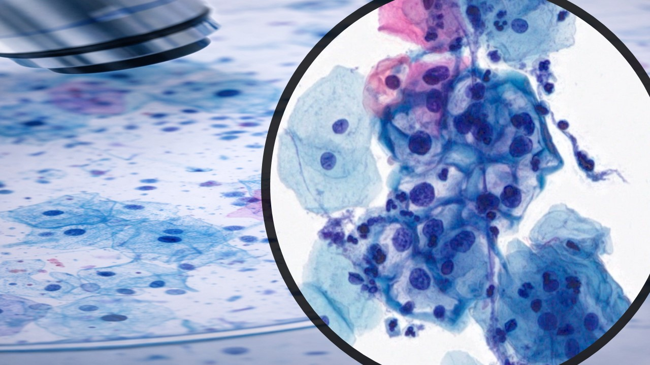
ALPHA PROLIPSIS
SKIN LESIONS FNA
Fine needle biopsy (FNB) and cytodiagnosis has found only limited application in primary tumors of the skin and subcutis due to the ease of surgical biopsy and of excision. The main indications for FNB are rapid, non-invasive investigation of suspected metastatic malignancy, and distinction between neoplasia and a reactive process likely to resolve spontaneously or respond to conservative treatment.
In patients with known malignancy, the nature of any nodules or thickenings related to surgical scars or elsewhere in the skin or subcutis the distinction can easily be made between suture granuloma, infection or other reactive process and recurrent or metastatic tumor. The possibility of a second primary or of de-differentiation of the original tumor can also be decided by cytology.
Cytological features
Some knowledge of the cytological features of skin adnexal tumors is needed to avoid mistaking these for metastatic malignancies. With some exceptions, a type-specific diagnosis of primary adnexal tumors by FNB is difficult or impossible but is not often of practical importance. From a clinical point of view, the information sought is mainly if the lesion is benign or malignant, and if malignant whether it is primary or metastatic. Cytology as a supplement to history and clinical findings can be useful in the management of patients with skin tumors.
Incisional biopsy or punch biopsy of pigmented lesions is generally contraindicated in view of the risk of spreading melanoma and of interfering with the histological diagnosis and staging on subsequent excision. Although not proven, FNB could have similar adverse effects and is therefore not recommended for localized pigmented skin lesions. However, since pigmented lesions are occasionally examined by FNB, cytopathologists must be familiar with the cytomorphology of melanocytic skin lesions (see below).
Scrape smears from tumors involving the epidermis or a mucous membrane is an alternative to biopsy in some cases, e.g. to confirm suspected squamous cell carcinoma, basal cell carcinoma or Paget's disease. Scrape smears can also be used to confirm advanced ulcerated malignant melanoma.
Dermatologists seeking an alternative treatment to surgical excision of basal cell carcinoma have shown some interest in cytological confirmation of this diagnosis.
Subcutaneous lipoma is one of the commonest 'tumors' seen in a community-based cytology practice. It may seem unnecessary to subject these clinically characteristic lesions to biopsy, but the patient and sometimes the referring doctor are often concerned and demand immediate reassurance. We have had some surprise findings in such cases and feel that FNB is generally justified. One example of several: a small, discrete, soft nodule was incidentally felt in the lower anterior abdominal wall of a 45-year-old woman. The clinical diagnosis was lipoma, but cytology showed a highly cellular, sarcomatous tumor, histologically confirmed as synovial sarcoma.
In general, FNB is not useful in the investigation of inflammatory skin disease except to provide material for microbiology, for example in tuberculosis and leprosy.
Health Tips
Accuracy of diagnosis
Only a few series of FNB of primary skin tumors with histo-logic correlation have been reported In one of the largest series, 89% of primary skin tumors were correctly diagnosed as benign or malignant, and specific typing was possible in 81% of cases.4Some skin adnexal tumors, in particular pilomatrixoma, have been reported as diagnostic pitfalls and a possible cause of false-positive cytological diagnosis.9Clinical correlation is essential.
Complications
The hypothetical risk of tumor cell seeding and of compro-mising subsequent histological examination as a result of FNB has been mentioned. Other significant complications have not been reported.
Technical considerations
We generally use 25- or 27-gauge needles and the non-aspiration technique for skin tumors, 23-25-gauge for sub-cutaneous lesions. Local anesthesia is rarely necessary. Two to four passes are usually required to obtain sufficient mate-rial and to compensate for possible tumor heterogeneity. Alcohol-fixed H&E- or Pap-stained smears and air-dried smears stained with Diff-Quik or MGG should be used in parallel. FNB of skin lesions often yield cohesive tissue frag-ments. Alcohol-fixation makes tissue fragments transparent whereas cell detail is poorly seen in fragments in air-dried smears. Squamous differentiation is more easily recognized in Pap-stained smears. On the other hand, air-dried MGG smears provide more information on secretory products and stromal components.
Thin superficial skin lesions, particularly if eroded or ulcer-ated, are best sampled by scraping cells off the surface. Keratin, crust and inflammatory exudate must be removed to ensure that intact, well-preserved cells are obtained from as deeply into the lesion as possible. Scraping with a sterile scalpel blade held at a blunt angle until the lesion bleeds slightly is recommended.10Wooden spatulas or other soft materials are less suitable since they tend to absorb the fluid component. FNB can be successful in some thin lesions using a 25-27-gauge needle inserted tangentially.