Alpha Prolipsis' Medical Services
Your reports will be available within 24 hours of receiving the sample
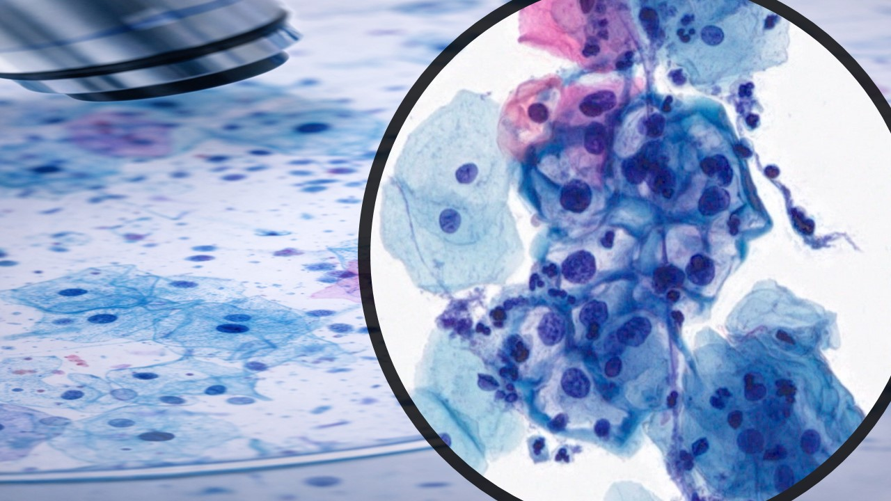
ALPHA PROLIPSIS
FINE NEEDLE ASPIRATION CYTOLOGY
Fine Needle Aspiration Biopsies (FNA, FNAC, FNAB)
Fine-needle aspiration (FNA)
Fine-needle aspiration (FNA) is a diagnostic procedure used to investigate lumps or masses. In this technique, a thin (23-25 gauge), hollow needle is inserted into the mass for sampling of cells that, after being stained, will be examined under a microscope (biopsy). The sampling and biopsy considered together are called fine-needle aspiration biopsy (FNAB) or fine-needle aspiration cytology (FNAC) (the latter to emphasize that any aspiration biopsy involves cytopathology not histopathology). Fine-needle aspiration biopsies are very safe, minor surgical procedures. Often, a major surgical (excisional or open) biopsy can be avoided by performing a needle aspiration biopsy instead. In 1981, the first fine-needle aspiration biopsy in the United States was done atMaimonides Medical Center, eliminating the need for surgery and hospitalization. Today, this procedure is widely used in the diagnosis of cancer and inflammatory conditions.
A needle aspiration biopsy is safer and less traumatic than an open surgical biopsy, and significant complications are usually rare, depending on the body site. Common complications include bruising and soreness. There is a risk, because the biopsy is very small (only a few cells), that the problematic cells will be missed, resulting in a false negative result. There is also a risk that the cells taken will not enable a definitive diagnosis.
For FNAs performed during normal business hours, the Cytology Department may be contacted to assist in the preparation of the smears. Contact the Cytology Laboratory for further information.
Health Tips
Fine Needle Aspiration: When It's Used
A fine needle aspiration is most often done on swellings or lumps located just under the skin.
A lump may be felt during a doctor's examination. Or it may be discovered on an imaging test such as:
• CT scan
• mammogram
• ultrasound
Imaging tests may also discover abnormal spots deeper inside the body.
Doctors may recommend fine needle aspiration for areas such as:
• cysts (fluid-filled lumps)
• nodules or masses (solid lumps)
• enlarged lymph nodes
Without a biopsy, it's usually hard for a doctor to confirm what these abnormal areas contain. And you may not know if they are a threat to your health.
The most common reason to get a fine needle aspiration is to test for cancer.
Most fine needle aspirations are done on these areas:
• breast
• thyroid gland
• lymph nodes in the neck, groin, or armpit
Those types of fine needle aspirations are performed through the skin.
Using endoscopy, doctors can also reach areas deeper in the body. An endoscopy uses a flexible tube with a light and camera attached. During an endoscopy, a doctor can do a fine needle aspiration on certain abnormal spots in the chest or abdomen.
Procedure of fine needle aspirations performed by clinicians
1. Determine the gross characteristics of the mass to be aspirated including location relative to other structures, estimated depth, consistency, and any evidence of pulsation or bruit.
2. Assemble the syringe and syringe pistol with attached needle (25 -23gauge).
3. Layout several glass slides, fixative and rinsing solution (Cytolyt or RPMI).
4. Label slides (using pencil), fixative and rinsing solution containers with patients name.
5. Check the requisition form if properly filled out - patient's name and Medical record number, attending full name, clinical information, site of aspiration.
6. Clean the skin over the puncture site with an alcohol skin preparation pad.
7. Grasp the mass firmly with the free hand and insert the needle in one swift motion.
8. Apply full vacuum pressure to the syringe with the pistol finger.
9. Move the needle back and forth within the mass at slightly different angles while full pressure is maintained.
10. Observe the junction of the needle and syringe for the appearance of any sample or continue to make back-and-forth passes within the mass about a dozen times.
11. Conclude the aspiration if the hub of a needle is filled with a material or after about dozen passes by releasing the trigger of the syringe pistol. DO NOT HOLD PRESSURE WHILE WITHDRAWING! If vacuum pressure is applied while withdrawing the needle, the sample will be pulled into the syringe where it will dry and be difficult to retrieve.
12. Withdraw the needle from the mass and place pressure on the puncture site with a sterile gauze pad.
13. Detach the needle from the syringe, fill the syringe with air and reattach the needle.
14. Express a small drop of aspirated material on one slide and gently spread the material by using a second slide.
15. Immediately fix one slide by dropping it into 95% alcohol. The second slide leave for air¬ drying.
16. The remaining material, express into a rinsing solution. Rinse the syringe well.
17. Stain air-dried slides with Diff Quik.
18. Perform additional pass/passes until adequate material is obtained.
19. Bring all material (slides and rinsing solution) to the cytology laboratory for processing.
20. If the aspirated lesion is cystic, aspirate as much fluid as possible and place in a clean, leak - proof rigid, labeled container.
21. Reaspirate any residual mass or a wall of a cyst and prepare smears as directed above.
What is a Fine Needle Aspiration Biopsy?
A fine needle aspiration (FNA) is a biopsy technique used to sample potentially abnormal tissue. This technique can be used for patients with either superficial or deep nodules. If the lesion is deeper and cannot be felt, imaging techniques, such as an ultrasound or CT, can be used to localize the lesion for FNA. Using sterile technique, a small needle is introduced into the mass and quickly moved back and forth to remove cells. This may be performed multiple times. The cells are placed on glass slides, stained, and examined under a microscope in order to provide a diagnosis. The pathologist or cytotechnologist may review the glass slides at the time of the procedure to decide whether or not additional samples are needed [this is called a rapid on-site adequacy assessment]. Depending on the type of cells present, additional tests may be performed to determine the final diagnosis.
Will the test spread the disease and are there any complications?
Because such a thin needle is used in this procedure the risk of spreading tumor essentially does not exist. Significant complications are extremely rare with this technique. The most common complication is for blood to collect at the site of the aspiration causing a hematoma (bruise), but this doesn’t usually require any treatment or at most you may want to apply a cold compress to the site. Even patients taking aspirin, Advil® or other non-steroidal anti-inflmmatory drugs should not experience significant bleeding. If you have a severe bleeding disorder or are taking blood thinners such as Heparin, Coumadin® or Plavix® you should discuss with their physician discontinuing the use of these drugs 3-5 days prior to the procedure. In the very rare event that you should experience significant difficulty in breathing some time after the procedure you should contact your physician.
How long will the procedure take and will I experience any discomfort?
The procedure takes about 15-20 minutes. Since such a small needle is used, most patients do not find this procedure very uncomfortable. In fact, we do not usually use a local anesthetic because most patients tell us that receiving the local anesthetic stings more than the procedure itself. Also use of a local anesthetic may mix with and distort the material we are trying to examine. If you experience discomfort after the procedure Aspirin or Tylenol or the application of an ice pack may help relieve any discomfort.
Will I need to take off from work?
You need to take off only the time required to have the procedure performed, you may go back to work or school the same day. If you need a note for your absence for your employer or school please notify our receptionist.
Can I eat or take medications before the procedure?
Yes, you do not need to alter your daily routine for this procedure.
Can I drive after the procedure? Are there any restrictions to my activity following the procedure?
Driving is fine. There are no restrictions to your normal activities.
When can I expect results from this test?
Your physician will generally receive results from this test within two working days. They may, however, be awaiting results from other tests before determining what is the best course of action for treatment. If you have not heard from your physician within a week you should contact them to determine what the plan of action may be.
What are the advantages of FNA?
FNA is a rapid procedure with minimal discomfort for the patient. The actual procedure takes only 10-20 minutes, but the entire process can take up to an hour or more if imaging or rapid on-site adequacy is necessary. Rapid on-site adequacy assessment reduces the chance that the biopsy will need to be repeated by ensuring that diagnostic material is present. This may save the patient an additional trip back to the doctor’s office or hospital. Since FNA is minimally invasive and it uses a very small needle, it is generally well-tolerated and typically requires little recuperation following the procedure. Results are usually available within a few days.
Who performs the FNA?
FNA is usually performed by a trained physician. Rapid on-site adequacy assessment, if available, is performed by a pathologist or certified cytotechnologist. All of the slides and additional test results, if necessary, are examined by a pathologist, who gives a final diagnosis. This information is typically written in a report and given to the treating physician, who will inform the patient of the findings.
What do I have to do to prepare for an FNA?
If the site to be biopsied is superficial (can be felt), no preparation is needed. Discuss with your doctor beforehand if you are taking aspirin or other blood thinners, have a history of a bleeding disorder, or have low platelets. If the nodule or mass is deep and requires imaging to be visualized (for example, in the lung), you will receive pre-procedure instructions either from radiology or the team performing the FNA.
Is an FNA painful?
One of the advantages of FNA is that it is a relatively pain-free procedure. For superficial lesions, anesthetic (such as lidocaine) is typically not necessary. The needle used is smaller than the needle used to draw blood. Patients feel a “poke” during the initial entrance of the needle after which a sensation of pressure is most commonly described. Biopsies of deeper locations, visualized by imaging techniques, may require sedation and/or anesthetic. Sedation is not indicated for FNAs of superficial or palpable (‘feel-able’) masses performed in the clinic or doctor’s office.
How accurate are the results from an FNA?
FNA is a quick and accurate way to determine the cause of the abnormal nodule or mass. Possible causes include infectious, inflammatory, and cancerous diseases. Often the FNA diagnosis will provide all of the information the clinician needs to determine treatment. In some cases which are usually related to the nature of disease, further diagnostic testing, either by repeat FNA or other tissue sampling technique, may be necessary.
Are there any complications of FNA?
Any time the skin is broken there is a chance of infection. To minimize this risk, the skin is cleaned prior to the procedure and sterile needles and syringes are used for each sampling pass. Bleeding, usually very minimal, is controlled with pressure after the needle is removed. A Band-Aid is usually placed over the FNA site when the procedure is finished. Slight bruising and mild soreness afterward are common. After the procedure, the patient can return to their normal activities without any restrictions. As with any health concern, it is best to call your doctor to report any unexpected post-procedure developments, such as moderate to severe pain, swelling, heat, or numbness, and to proceed to the emergency department if symptoms are severe.
What are FNAs and core biopsies for?
Sometimes a small sample of breast cells or breast tissue may be taken from the breast to help make a diagnosis. This is most commonly done using a core biopsy. Sometimes a fine needle aspiration (FNA) or another procedure, such as a punch biopsy, may be used. The sample is then sent to the laboratory where it is looked at under a microscope.
A mammogram or ultrasound may be used as a guide to pinpoint the area before the sample is taken, particularly when it’s very small or cannot be felt.
If you’re taking aspirin or any anticoagulants (blood-thinning tablets), let the doctor know before having a core biopsy or an FNA.
Having a core biopsy or an FNA doesn’t necessarily mean you have breast cancer.
Core biopsy (also called core needle biopsy)
A core biopsy uses a hollow needle to take one or more samples of breast tissue from the area of concern. Because tissue is taken rather than cells, it gives more detailed information.
After local anaesthetic is given to numb the area, a small cut is made in the skin so that samples of tissue can be taken. Sometimes you might be asked to lie on your front while this is done. The tissue is then sent to the laboratory where it’s examined under a microscope to establish a diagnosis.
If the area of concern can only be seen on a mammogram, you may have a stereotactic core biopsy. This is where a sample of tissue is taken using a needle biopsy device connected to a mammogram machine and linked to a computer. This helps locate the exact position of the area to be biopsied. Images of the breast are taken from two different angles to help guide the needle to the precise location. You will be given a local anaesthetic and will be in a sitting position or lying down on a specialised examination couch. It may feel a little uncomfortable as the mammogram plates are pressed onto the breast throughout.
Whichever way the core biopsy is done, a small dressing or a plaster will usually be applied, and you’ll be asked to keep this on for a day or so afterwards. Sometimes very thin strips of adhesive tape are used to help the edges of the wound to close. Once the local anaesthetic wears off, your breast may ache and may also become bruised. You can take pain relief if the area is tender or painful. You’ll be given more information about this before you leave the clinic.
What is Ultrasound-Guided Fine Needle Aspiration Biopsy of the Thyroid?
During a fine needle aspiration biopsy of the thyroid, a small sample of tissue is removed from the thyroid gland. The thyroid gland is located in front of the neck just above the neckline and is shaped like a butterfly, with two lobes on either side of the neck connected by a narrow band of tissue.
Nodules or abnormalities in the body are often detected by imaging examinations. However, it is not always possible to tell from these imaging tests whether a nodule is benign (non-cancerous) or cancerous.
A needle biopsy, also called a needle aspiration, involves removing some cells—in a less invasive procedure involving a hollow needle—from a suspicious area within the body and examining them under a microscope to determine a diagnosis.
What are some common uses of the procedure?
hyroid biopsy is used to find the cause of a nodule in the thyroid gland.
When a nodule is detected, imaging tests may be performed to help determine if it is benign (non-cancerous) or malignant (cancerous). If imaging studies cannot clearly define the abnormality, a biopsy may be necessary.
How should I prepare?
Please notify your physician if you are taking any blood thinning agents, such as aspirin, Lovenox®, Plavix® or Coumadin®.
Usually, no special preparations are required for this procedure.
For biopsies performed in children, sedation may be used. Specific instructions will be given at the time of scheduling.
What does the equipment look like?
The needle used is a thin, fine-gauge needle that is smaller in diameter than the needle used in most blood draws (usually a 25 or 27 gauge 1.5 inch needle). The aspiration may be done with a needle or with a needle that is attached to a syringe. The syringe may be in a plastic or metal holder to make it easier for the doctor to aspirate the cells.
Ultrasound is used to guide accurate placement of the needle within the thyroid nodule.
Ultrasound scanners consist of a console containing a computer and electronics, a video display screen and a transducer that is used to do the scanning. The transducer is a small hand-held device that resembles a microphone, attached to the scanner by a cord. Some exams may use different transducers (with different capabilities) during a single exam. The transducer sends out high-frequency sound waves (that the human ear cannot hear) into the body and then listens for the returning echoes from the tissues in the body. The principles are similar to sonar used by boats and submarines.
The ultrasound image is immediately visible on a video display screen that looks like a computer or television monitor. The image is created based on the amplitude (loudness), frequency (pitch) and time it takes for the ultrasound signal to return from the area within the patient that is being examined to the transducer (the device placed on the patient's skin to send and receive the returning sound waves), as well as the type of body structure and composition of body tissue through which the sound travels. A small amount of gel is put on the skin to allow the sound waves to travel from the transducer to the examined area within the body and then back again. Ultrasound is an excellent modality for some areas of the body while other areas, especially air-filled lungs, are poorly suited for ultrasound.
How does the procedure work?
The physician inserts a fine gauge needle through the skin and advances it into the thyroid nodule.
Samples of the cells are then obtained and put on a slide for review by the pathologist.
How is the procedure performed?
Image-guided, minimally invasive procedures such as fine needle aspiration of the thyroid are most often performed by a specially trained radiologist with experience in needle aspiration and ultrasound.
Needle biopsies are usually done on an outpatient basis.
The neck will be cleansed with antiseptic. Medicine to numb the area may or may not be used. An ultrasound transducer with a small amount of sterile water soluble gel will be placed on your neck over the thyroid nodule. The radiologist will insert the needle through the skin under direct imaging guidance, advance it to the site of the thyroid nodule and aspirate samples of tissue. After the sampling, the needle will be removed. New needles will be reinserted if additional samples are required. Several specimens may be needed for a complete analysis.
Once the biopsy is complete, pressure will be applied to the area to decrease the risk of bleeding. A bandage may be placed if necessary. No sutures are needed.
This procedure is usually completed in less than 30 minutes.
What will I experience during and after the procedure?
During the test, you will lie on your back with a pillow under your shoulders, your head tipped backward, and your neck extended. This position makes it easier for the radiologist to access the thyroid gland.
You may feel some pressure on your neck from the ultrasound transducer and mild discomfort as the needle is moved to obtain the cells.
You will be asked to remain still and not to cough, talk, swallow or make a sound during the procedure.
Aftercare instructions vary, but generally you can resume normal activities and any bandage can be removed within a few hours.
The biopsy site may be sore and tender for one to two days. You may take nonprescription pain medicine, such as acetaminophen, to relieve any discomfort.
Who interprets the results and how do I get them?
A pathologist examines the removed specimen and makes a final diagnosis so that treatment planning can begin. Depending on the facility, the radiologist or your referring physician will discuss the results with you.
What are the benefits vs. risks?
Benefits
The results of needle biopsy of the thyroid are close to 95% accurate for adequate biopsies.
Needle biopsy is a reliable method of obtaining tissue samples that can help diagnose whether a nodule is benign (non-cancerous) or malignant.
A needle biopsy is less invasive than open and closed surgical biopsies, both of which involve a larger incision in the skin and local or general anesthesia.
Generally, the procedure is not painful and the results are as accurate as when a tissue sample is removed surgically.
Recovery time is brief and patients can soon resume their usual activities.
Risks
Bleeding at the site of biopsy.
Infection.
Injury to structures adjacent to the thyroid.
What are the limitations of Needle Biopsy of the Thyroid?
Complications of thyroid biopsy are rare since the procedure is done under direct imaging guidance and with a fine needle.
In some cases, the procedure may have to be repeated in order to obtain diagnostic results.
Fine needle aspiration biopsy facts
Fine needle aspiration biopsy (FNAB) of the thyroid is a procedure used to detect cancer in a thyroid nodule or to treat thyroid cysts.
The chance that a thyroid nodule is malignant varies with age, gender, radiation exposure, and other factors.
fine needle aspiration biopsy is performed in a doctor's office and takes about 20 minutes.
Complications are rare, but include bleeding, bruising, and infection.
Results help determine further management and treatment and are usually available within a week.
What are thyroid nodules?
The thyroid gland is found in the neck just below the "Adam's apple." This gland is responsible for producing thyroid hormone, which is an important hormone that stimulates the metabolism of the body. Thyroid nodules are so common that up to half of all people have one, without any symptoms or effect. Like many things, the thyroid gland gets "lumpier" as we get older and the frequency of these nodules increases with age. In fact, many are found incidentally during routine examinations or radiology testing. Thyroid nodules are also more common in women than in men. Interestingly, because women have so many more nodules than men, the incidence of detected cancer is higher in women than in men by virtue of absolute numbers. However, each individual nodule is more likely to be cancerous if found in a man.
Doctors always hold a degree of concern whenever a new growth is detected on the body, regardless of the tissue involved. The concern is whether or not the growth or nodule is cancer. Fortunately, fewer than 10% of thyroid nodules are malignant. The majority of thyroid nodules are harmless growths, known as adenomas, and are contained within a capsule. Even though cancerous nodules are uncommon, the doctor will take the necessary measures to be certain.
What is the initial assessment of a thyroid nodule?
All patients with a thyroid nodule should undergo a complete medical history and physical examination. Specific questions regarding the onset of the nodule, related pain or discomfort, symptoms of thyroid disease, and family history are addressed. In addition, the doctor will take into account the patient's age and sex when evaluating the possibility of malignancy. Patients with a history of head and neck radiation (which was commonly used in the 1950's as an acne treatment) are at a higher risk. Cancerous nodules are also more frequent in men as compared to women. The doctor will also look for general symptoms of thyroid disease in addition to other illnesses. The size and characteristics of the nodule are assessed. Is it soft or firm? Does it move with swallowing, or is it fixed? Is there more than one nodule? Are there other nodes involved? Does it hurt when the nodule is touched? The answers to these questions will help the doctor evaluate what further investigations, if any, are necessary.
The following is a list of factors that increase the suspicion of malignancy:
Age: Patients less than 30 years of age and greater than 60 years of age have a higher risk of cancer in a thyroid nodule as do children;
Associated symptoms such as difficulty swallowing or hoarseness;
History of head and neck irradiation;
A hard, fixed nodule on examination;
Surrounding enlarged lymph nodes; and
Previous history of thyroid cancer in the family.
Nodules are less concerning to a physician if it is one of many present in the gland, and also if the nodule is hyperfunctioning (or "hot") using nuclear thyroid imaging.
After the initial evaluation, the doctor may choose to order thyroid blood tests or imaging scans to determine the functional activity of a thyroid nodule and its anatomy. The cornerstone in the assessment of a solitary thyroid nodule is a procedure known as fine needle aspiration biopsy ("FNAB") of the thyroid gland.
Fine needle aspiration biopsy (FNAB) of the thyroid gland; why is it done?
View the Hyperthyroidism Slideshow Pictures
Hyperthyroidism Slideshow Pictures
Take the Thyroid Disorder Quiz
Thyroid Symptoms and Solutions Slideshow Pictures
Related Diseases
Images & Quizzes
Index
Hyperthyroidism Slideshow Pictures
Take the Thyroid Disorder Quiz
Thyroid Symptoms and Solutions Slideshow Pictures
Patient Comments: Fine-Needle Aspiration Biopsy of the Thyroid - Experience
Patient Comments: Fine Needle Aspiration Biopsy - Complications
Patient Comments: Fine Needle Aspiration Biopsy - Results
Fine needle aspiration biopsy facts
What are thyroid nodules?
What is the initial assessment of a thyroid nodule?
Fine needle aspiration biopsy (FNAB) of the thyroid gland; why is it done?
Should fine needle aspiration biopsy be done on all thyroid nodules?
How is fine needle aspiration biopsy performed?
What are the complications of fine needle aspiration biopsy of the thyroid?
What happens to the thyroid tissue obtained at the fine needle aspiration biopsy?
Summary
Fine needle aspiration biopsy facts
Fine needle aspiration biopsy (FNAB) of the thyroid is a procedure used to detect cancer in a thyroid nodule or to treat thyroid cysts.
The chance that a thyroid nodule is malignant varies with age, gender, radiation exposure, and other factors.
fine needle aspiration biopsy is performed in a doctor's office and takes about 20 minutes.
Complications are rare, but include bleeding, bruising, and infection.
Results help determine further management and treatment and are usually available within a week.
What are thyroid nodules?
The thyroid gland is found in the neck just below the "Adam's apple." This gland is responsible for producing thyroid hormone, which is an important hormone that stimulates the metabolism of the body. Thyroid nodules are so common that up to half of all people have one, without any symptoms or effect. Like many things, the thyroid gland gets "lumpier" as we get older and the frequency of these nodules increases with age. In fact, many are found incidentally during routine examinations or radiology testing. Thyroid nodules are also more common in women than in men. Interestingly, because women have so many more nodules than men, the incidence of detected cancer is higher in women than in men by virtue of absolute numbers. However, each individual nodule is more likely to be cancerous if found in a man.
Doctors always hold a degree of concern whenever a new growth is detected on the body, regardless of the tissue involved. The concern is whether or not the growth or nodule is cancer. Fortunately, fewer than 10% of thyroid nodules are malignant. The majority of thyroid nodules are harmless growths, known as adenomas, and are contained within a capsule. Even though cancerous nodules are uncommon, the doctor will take the necessary measures to be certain.
Quick Guide
Thyroid Problems Explained
Thyroid Problems Explained
What is the initial assessment of a thyroid nodule?
All patients with a thyroid nodule should undergo a complete medical history and physical examination. Specific questions regarding the onset of the nodule, related pain or discomfort, symptoms of thyroid disease, and family history are addressed. In addition, the doctor will take into account the patient's age and sex when evaluating the possibility of malignancy. Patients with a history of head and neck radiation (which was commonly used in the 1950's as an acne treatment) are at a higher risk. Cancerous nodules are also more frequent in men as compared to women. The doctor will also look for general symptoms of thyroid disease in addition to other illnesses. The size and characteristics of the nodule are assessed. Is it soft or firm? Does it move with swallowing, or is it fixed? Is there more than one nodule? Are there other nodes involved? Does it hurt when the nodule is touched? The answers to these questions will help the doctor evaluate what further investigations, if any, are necessary.
The following is a list of factors that increase the suspicion of malignancy:
Age: Patients less than 30 years of age and greater than 60 years of age have a higher risk of cancer in a thyroid nodule as do children;
Associated symptoms such as difficulty swallowing or hoarseness;
History of head and neck irradiation;
A hard, fixed nodule on examination;
Surrounding enlarged lymph nodes; and
Previous history of thyroid cancer in the family.
Nodules are less concerning to a physician if it is one of many present in the gland, and also if the nodule is hyperfunctioning (or "hot") using nuclear thyroid imaging.
After the initial evaluation, the doctor may choose to order thyroid blood tests or imaging scans to determine the functional activity of a thyroid nodule and its anatomy. The cornerstone in the assessment of a solitary thyroid nodule is a procedure known as fine needle aspiration biopsy ("FNAB") of the thyroid gland.
Fine needle aspiration biopsy (FNAB) of the thyroid gland; why is it done?
Readers Comments 100 Share Your Story
A biopsy to obtain tissue for analysis is the best technique for detecting or ruling out the presence of cancer. For many years, a core biopsy of the thyroid was the procedure of choice. This method involved a large biopsy, which was often more difficult for patients. fine needle aspiration biopsy has now become the method of choice for obtaining samples of thyroid tissue. The procedure is technically quite simple. When performed properly, the testing has a false negative rate of less than 5%. This means that a positive finding, such as cancer, will be missed fewer than five times out of 100.
The fine needle aspiration is also performed to treat thyroid cysts. A thyroid cyst is a fluid-filled sac within the thyroid gland. Aspiration of the cyst with a needle and syringe can shrink the swelling from the cyst and the fluid removed can be analyzed for cancer.
Should fine needle aspiration biopsy be done on all thyroid nodules?
There are certain situations in which your physician may elect not to perform a biopsy of a nodule. For example, in a patient with an over- active thyroid (hyperthyroidism), the chance for a nodule to be cancerous is significantly less, particularly if other studies (such as nuclear thyroid imaging) show that the nodule is producing thyroid hormone (a "hot" nodule).
A doctor may recommend fine needle aspiration biopsy of the thyroid in the following situations:
To make a diagnosis of a thyroid nodule;
To help select therapy for a thyroid nodule;
To drain a cyst that may be causing pain; or
To inject a medication to shrink a recurrent cyst.
How is fine needle aspiration biopsy performed?
In most cases, if the nodule can be felt, a biopsy can be performed in the doctors office. In some cases an ultrasound may be needed to help guide the biopsy, (for example, if the nodule cannot be felt without difficulty or if the nodule has areas within it that specifically should be biopsied).
Little preparation by the patient is required. There is no need to fast or to withhold medications on the day of the biopsy. Occasionally, though, a patient may be asked not to take blood thinning medication either before or on the day of the biopsy. After an examination to pinpoint the nodule, the patient is asked to lie down and the neck is exposed. Depending on the location of the nodule and the type of clothes the patient is wearing, he or she may be asked to change into a gown. The doctor drapes the area around the neck and cleans the neck off. This is usually done with iodine, which is a brown liquid that sterilizes the skin. Some doctors may choose to inject a local anesthetic. Often, the injection of the anesthetic results in an initial discomfort, like a bee sting. The majority of doctors who regularly perform fine needle aspiration biopsies of the thyroid do not use a local anesthetic for this reason. Since the needle used for fine needle aspiration biopsy is so fine, anesthesia often results in simply another uncomfortable poke for the patient. If a patient is particularly concerned and nervous, a topical anesthetic preparation may be applied, which takes 10 to 20 minutes to work, thus prolonging the procedure. A patient undergoing fine needle aspiration biopsy should discuss any preferences for local anesthetic before the procedure begins. Most patients undergoing fine needle aspiration biopsy forego the use of any anesthetic and do very well.
Once the patient is ready, a small, fine-gauge needle is inserted into the nodule. The needle is smaller in diameter than the needle used in most blood draws (usually a 25 gauge 1.5 inch needle). The patient holds his breath while the needle is rocked gently to obtain as much tissue as possible. (The reason for holding the breath is to minimize movement of the structures in the neck.) The needle is then withdrawn and pressure is applied over the thyroid area to minimize bleeding. This procedure is usually repeated four to six times to ensure that an adequate amount of tissue has been collected. After the procedure, pressure is applied over the neck area for 5 to 10 minutes to assure that the bleeding has stopped. The pressure also helps to reduce any swelling that may occur. The entire
procedure usually takes less than 20 minutes.
What are the complications of fine needle aspiration biopsy of the thyroid?
Most patients notice very little bleeding or swelling. There may be some discomfort in the area for a few hours after the biopsy, which is usually relieved with acetaminophen (Tylenol). Some patients like to put an ice pack over the area when they get home, but most do well without such measures. The risks of fine needle aspiration biopsy of the thyroid include bleeding, infection, and cyst formation, but these complications are exceedingly rare. Patients should contact their doctor if they notice any excessive bruising or swelling in the area of the biopsy, if they have persistent pain in the area, or if they develop a fever.
What happens to the thyroid tissue obtained at the fine needle aspiration biopsy?
After the procedure, the tissue obtained is prepared onto glass slides and sent to the pathologist for evaluation. First, the pathologist determines whether or not enough thyroid tissue has been obtained for analysis. (When there is an insufficient amount, a repeat fine needle aspiration biopsy is necessary.) After analysis, the tissue is classified. Although the classifications used by pathologists vary, the tissue is usually reported as (1) benign; (2) malignant; (3) suspicious; or (4) indeterminate. The chance of a false negative test (a test report that is negative when cancer is actually present) varies from 0-5%, depending on where the test is performed. The chance of a false positive (a test report showing cancer when there is no cancer present) is less than 5% and is usually due to the presence of degenerating cells or atypical cells. These results are reported back to the doctor's office, usually within one week. At this point, the doctor discusses the implications of the report and outlines further treatment, if needed based on the results.
Medication uses
This type of sampling is performed for one of two reasons:
A biopsy is performed on a lump or a tissue mass when its nature is in question.
For known tumors, this biopsy is performed to assess the effect of treatment or to obtain tissue for special studies.
When the lump can be felt, the biopsy is usually performed by a cytopathologist or a surgeon. In this case, the procedure is usually short and simple. Otherwise, it may be performed by an interventional radiologist, a doctor with training in performing such biopsies under x-ray or ultrasound guidance. In this case, the procedure may require more extensive preparation and take more time to perform.
Also, fine-needle aspiration is the main method used for chorionic villus sampling, as well as for many types of body fluid sampling.
It is also used for ultrasound-guided aspiration of breast abscess,[3] of breast cysts, and of seromas.
Preparation
Several preparations may be necessary before this procedure.
No use of aspirin or non-steroidal anti-inflammatory medications (e.g. ibuprofen, naproxen) for one week before the procedure;
No food intake a few hours before the procedure;
Routine blood tests (including clotting profile) must be completed two weeks before the biopsy;
Suspension of blood anticoagulant medications;
Antibiotic prophylaxis may be instituted.
Before the procedure is started, vital signs (pulse, blood pressure, temperature, etc.) may be taken. Then, depending on the nature of the biopsy, an intravenous line may be placed. Very anxious patients may want to be given sedation through this line. For patients with less anxiety, oral medication (Valium) can be prescribed to be taken before the procedure.
Procedure
The skin above the area to be biopsied is swabbed with an antiseptic solution and draped with sterile surgical towels. The skin, underlying fat, and muscle may be numbed with a local anesthetic, although this is often not necessary with superficial masses. After locating the mass for biopsy, using x-rays or palpation, a special needle of very fine diameter is passed into the mass. The needle may be inserted and withdrawn several times. There are many reasons for this:
One needle may be used as a guide, with the other needles placed along it to achieve a more precise position.
Sometimes, several passes may be needed to obtain enough cells for the intricate tests which the cytopathologists perform.
After the needles are placed into the mass, cells are withdrawn by aspiration with a syringe and spread on a glass slide. The patient's vital signs are taken again, and the patient is removed to an observation area for about 3 to 5 hours.
For biopsies in the breast, ultrasound-guided fine needle biopsy is the most common.The biopsy is advised.
Post-operative care and complications
As with any surgical procedure, complications are possible, but major complications due to thin needle aspiration biopsies are fairly uncommon, and when complications do occur, they are generally mild. The kind and severity of complications depend on the organs from which a biopsy is taken or the organs gone through to obtain cells.
After the procedure, mild analgesics are used to control post-operative pain. Aspirin or aspirin substitutes should not be taken for 48 hours after the procedure (unless aspirin is prescribed for a cardiac or neurological condition). Since sterility is maintained throughout the procedure, infection is rare. But should an infection occur, it will be treated with antibiotics. Bleeding is the most common complication of this procedure. A slight bruise may also appear. If a lung or kidney biopsy has been performed, it is very common to see a small amount of blood in sputum or urine after the procedure. Only a small amount of bleeding should occur. During the observation period after the procedure, bleeding should decrease over time. If more bleeding occurs, this will be monitored until it subsides. Rarely, major surgery will be necessary to stop the bleeding.
Other complications depend upon the body part on which the biopsy takes place:
Lung biopsies are frequently complicated by pneumothorax (collapsed lung). This complication can also accompany biopsies in the upper abdomen near the base of the lung. About one-quarter to one-half of patients having lung biopsies will develop pneumothorax. Usually, the degree of collapse is small and resolves on its own without treatment. A small percentage of patients will develop a pneumothorax serious enough to require hospitalization and placement of a chest tube for treatment. Although it is impossible to predict in whom this will occur, collapsed lungs are more frequent and more serious in patients with severe emphysema and in patients in whom the biopsy is difficult to perform.
For biopsies of the liver, bile leakages may occur, but these are quite rare.
Pancreatitis (inflammation of the pancreas) may occur after biopsies in the area around the pancreas.
In biopsies in the area of the breast, bleeding and bruising may occur, less frequently also infection (rarely) or (very rarely, and only if performed near the chest wall) pneumothorax.
Deaths have been reported from needle aspiration biopsies, but such outcomes are extremely rare.