Alpha Prolipsis' Medical Services
Your reports will be available within 24 hours of receiving the sample
ALPHA PROLIPSIS
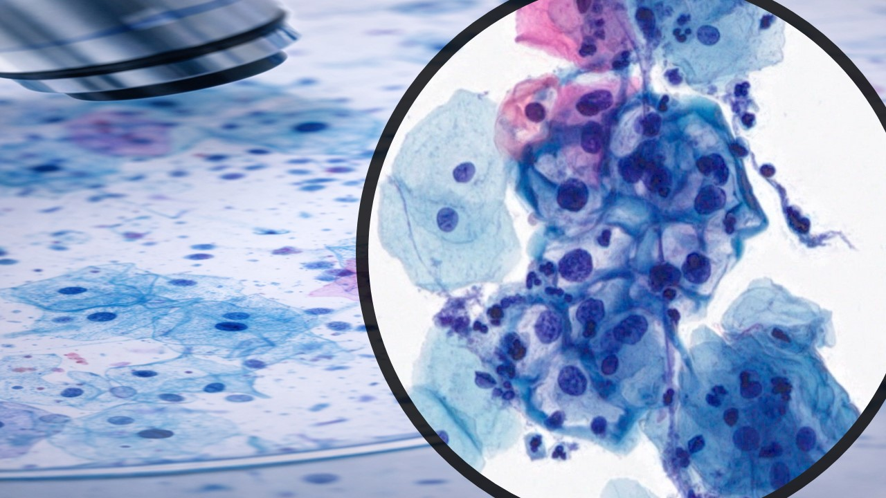
SALIVARY GLANDS FNA
Our goal is to provide accurate laboratory testing, with a reasonable turnaround time, in a cost-effective manner.
Fine needle aspiration cytology (FNAC)
Fine needle aspiration cytology (FNAC) has become popular as a valuable tool in preoperative assessment of breast masses, and it shows high accuracy, sensitivity, and specificity. It has gained popularity due to its fast and easy approach, being inexpensive, and can be performed with little complications. To differentiate benign from malignant lesions is one of the major goals of FNAC.
Major salivary glands are easily accessible; therefore, they are optimal targets for fine-needle aspiration (FNA). This technique has been used for decades in Europe, but it is still controversial in the United States. The large numbers of benign and malignant tumors that can originate in the glands, as well as the lack of cytological markers to accurately diagnose the various tumors, often foster uncertainty in fine-needle aspiration (FNA) diagnosis of salivary gland masses. Therefore, the primary challenge of fine-needle aspiration (FNA) is differentiating benign from malignant disease and then differentiating the various malignancies.
Fine-needle aspiration (FNA) enables high accuracy in identifying the nature of a lesion and can provide the following information:
Differentiation of inflammatory from neoplastic disease: This is especially important in patients who are immunosuppressed.
Culture for suspected infectious masses
Differentiation of benign from malignant disease
Differentiation of the specific tumor cell type
Determination of site of origin, ie, whether the tumor has arisen from the parotid or if it is metastatic
Squamous cell carcinoma evaluation: Squamous cell carcinoma can be accurately diagnosed, and treatment can be planned based on fine-needle aspiration (FNA) findings. A highly cellular mucoepidermoid carcinoma may appear to be squamous cell carcinoma by fine-needle aspiration (FNA) cytology. This difference is purely academic and does not change the treatment.
Malignancy determination: The pathologist is usually able to very accurately render an opinion about the malignant or benign nature of the lesion. This is a great help in promptly guiding treatment, and this is further addressed below.
The use of fine-needle aspiration (FNA) allows for immediate assessment of the lesion in an outpatient setting.
In the author's institution, most fine-needle aspirations (FNAs) of the salivary glands are performed in an outpatient setting.
Immediate assessment of the material is usually possible within 15 minutes.
This initial assessment is helpful in relieving the anxiety of the patient and aids in clinical decision-making.
If the initial aspiration is not satisfactory or other studies are needed (eg, flow cytometry, lymphoma), additional aspirations are often necessary.
Fine-needle aspiration (FNA) of the salivary glands is a safe and reliable technique in the primary diagnosis and follow-up of patients. This technique is used with great success in Europe and is gaining increased popularity in the United States as the experience of pathologists and confidence of clinicians increase. With the application of special techniques, fine-needle aspiration (FNA) can be a very important part of clinical decision-making and preoperative counseling in most cases.
As always, the patient's overall status must be assessed, and an individual treatment plan must be created in each case. Although patients with high cardiac risk may be best served by observation given a benign fine-needle aspiration (FNA) diagnosis, young healthy patients may require surgical intervention regardless of a negative fine-needle aspiration (FNA) finding. A negative fine-needle aspiration (FNA) finding alone should not prevent surgical intervention when it is otherwise clinically indicated. Similarly, fine-needle aspiration (FNA) alone cannot dictate the extent of surgery because that judgment is based on tumor location and patient status.
Sacrifice of the facial nerve or other structures is generally best guided by findings at surgery rather than mindless obedience to preoperative dictums, as noted by Spiro: "Facial nerve preservation is ill advised when a parotid tumor has to be transected to spare the nerve. This rule applies even when the tumor is benign." This is not to say that decisions are always easy. On the contrary, making on-the-spot decisions with unusual tumor types that cannot be diagnosed with certainty at the time of surgery is often difficult.
Ultimately, diagnosis and treatment depend upon the experience of the pathologist and surgeon when dealing with anything less than permanent section diagnosis. In a center that has a relatively large volume and a pathologist with a great deal of experience and interest in salivary gland tumors, fine-needle aspiration (FNA) and/or frozen section diagnosis may be adequate to make preoperative and operative decisions.
Health Tips
Clinical implications
The use of fine-needle aspiration (FNA) in working up salivary gland tumors is also cost effective. Two recent studies have shown that the use of fine-needle aspiration (FNA) in salivary gland tumors is cost-effective and diagnostic. [5, 6] Specifically, Layfield et al demonstrated that the routine use of fine-needle aspiration (FNA) in the work-up of salivary gland lesions saves up to $70,000 per 100 patients. At the same time, fine-needle aspiration reduces the operative intervention by 65% in submandibular masses and by 35% in parotid masses.
Technique
Depending on the clinical situation, the cytopathologist, surgeon, or radiologist may perform the fine-needle aspiration (FNA). Identification of the nodule is essential to the successful aspiration. Deep lesions that are less than 1 cm in diameter are best aspirated under ultrasonographic or CT scanning guidance.
The skin is cleaned with an alcohol swab. A 25- or 23-gauge needle is used on a plastic disposable 10- or 20-mL syringe attached to a plastic or metal holder. The nodule is immobilized between the fingers, and the needle tip is rapidly directed through the skin into the nodule. Once the needle enters the mass, the needle is continuously aspirated while the needle is rapidly moved in and out to obtain the sample. Suction is then relieved, and the needle is withdrawn and detached from the syringe. Air is aspirated, and the material is expelled on glass slides (see the images below). The material is gently but rapidly smeared on the slides and immediately dipped in alcohol fixative. Some of the slides can be left to air dry for Diff-Quick staining and rapid evaluation. The fixed material is stained by Papanicolaou or hematoxylin-eosin stains, which are both excellent for nuclear detail.
If necessary, a cellblock is prepared from cells entrapped in a blood clot or tiny tissue particles obtained by fine-needle aspiration (FNA). A cellblock enables the pathologist to examine the tissue similarly to a biopsy, and multiple sections can be obtained from paraffin-embedded material for special studies (immunohistochemistry). The syringe is rinsed into a container with formalin for adequate fixation of cellblock material
Complications
Fine-needle aspirations (FNAs) are safely performed worldwide. The complications of salivary gland fine-needle aspirations (FNAs) are rare, but 6 potential complications exist, including the following:
Needle track contamination by malignant cells: Numerous studies indicate that needle tract contamination by malignant cells is a rare complication despite thousands of fine-needle aspirations (FNAs) performed worldwide. A positive correlation exists with number of passes and needle size.
Local hemorrhage: Because major salivary glands are in close proximity to the great vessels of the head and neck, local hemorrhage due to piercing of these vessels is possible but very unlikely. Physicians can prevent hematoma formation by applying firm pressure in the area of aspiration immediately after the procedure.
Infection: This risk is no greater than that of venipuncture and is closely correlated with the patient's immune status. Adherence to sterile techniques and cleaning the skin with alcohol minimizes this risk.
Warthin tumor (papillary cystadenoma lymphomatosum or adenolymphoma) has a high predisposition for parotitis from fine-needle aspiration (FNA) due to a combination of cystic spaces surrounded by oncocytic cells and poor blood supply.
Syncope: Some patients are prone to vasovagal reactions. Physicians should be prepared for this complication. Perform aspiration while the patient is lying down or sitting.
Dissemination of the dislodged tumor cells through lymphatics and blood vessels: This risk is certainly lower in fine-needle aspiration (FNA) than in incisional biopsy.
Contraindications
The general contraindications to fine-needle aspiration (FNA) include the following:
Bleeding diathesis: Because control of local hemorrhage depends on hemostasis and clotting, bleeding diathesis is a contraindication.
Uncooperative patient: Most nondiagnostic fine-needle aspiration (FNA) findings result from noncooperation by the patient. Prior to fine-needle aspiration (FNA), the physician should calm the apprehensive patient.
Skin infection in the area of fine-needle aspiration (FNA)
Fine-needle aspiration accuracy
Before making a definitive treatment decision, combining fine-needle aspiration (FNA) results with other data is prudent clinical practice. A typical example is a cytologic diagnosis of a lymphoepithelial cyst. The cyst is common in patients who have immunodeficiency virus (HIV); these patients tend to have bilateral lymphoepithelial cysts, and their findings are typical on CT scanning. Typically, these patients can be managed by serial examinations and occasional serial therapeutic aspiration. Lymphomas may also be accurately diagnosed if adequate tissue can be obtained for flow cytometry. A patient with a fine-needle aspiration (FNA) diagnosis of Warthin tumor may be best served by having a technetium scan confirmation without surgery if significant medical contraindications to surgery exist.
Recurrent tumors may also be accurately diagnosed using fine-needle aspiration (FNA). Some situations exist where a benign fine-needle aspiration (FNA) cytologic appearance should be viewed with suspicion. If the mass is painful, appears to be invading surrounding structures, or co-exists with an ipsilateral facial nerve palsy and the fine-needle aspiration (FNA) is benign, then the mass may be malignant despite a benign cytologic appearance.
Although fine-needle aspiration (FNA) is very accurate, situations exist where the final permanent pathology differs from the fine-needle aspiration (FNA) result. The following statistics demonstrate the high level of sensitivity and specificity of FNA in salivary gland lesions.
In 6249 cases of primary salivary gland lesions, fine-needle aspiration (FNA) biopsies revealed 4642 benign lesions and 1607 malignant lesions. The overall diagnostic accuracy of fine-needle aspiration (FNA) was 48%. Sensitivity for detecting malignant and benign tumors was 73% and 91%, respectively.
Benign lesions that were most often misdiagnosed as malignant were monomorphic adenoma (53% false positive rate), intraparotid lymph node (36%), oncocytoma (18%), and granulomatous sialadenitis (10%). The malignant lesions that were most often misdiagnosed as benign were lymphoma (57%), acinic cell carcinoma (49%), low-grade mucoepidermoid carcinoma (43%), and adenoid cystic carcinoma (33%).
Because of the referral pattern at these institutions, the distribution of the salivary gland lesions summarized here does not necessarily reflect the true incidence in the general population.
Pitfalls of diagnosis in salivary gland fine-needle aspiration (FNA)
Sampling error: Correct identification and immobilization of the lesion should allow the pathologist to avoid undersampling the lesion. Having the pathologist who performs the fine-needle aspiration (FNA) also interpret the lesions is a tremendous advantage.
Interpretation error: Pathologist experience minimizes this error.
Bias: Pathologist bias, augmented by the clinician's opinion, has been shown to lead to errors in diagnosis. [10]
Lack of flow cytometry use: Many of the false-positive and false-negative interpretations of lymphoma could have been prevented with flow cytometry.
Lack of immunophenotyping studies: These studies can improve interpretation of low-grade lymphoproliferative processes.
Technical problems: Air-drying is one of the most common technical problems. Romanowsky-type stains are superior to Papanicolaou stain for assessing stromal elements on salivary gland fine-needle aspirations (FNAs).
Role of special techniques
Some tumors have unique characteristics that can be discovered using special techniques. Although these are helpful, they are not always completely diagnostic.
Special stains
Mucin stains (eg, mucicarmine, periodic acid-Schiff [PAS]) can identify mucin in mucoepidermoid carcinomas.
PAS can identify the glycogen in acinic cell carcinomas.
Immunohistochemistry
S100: Some spindle cell myoepitheliomas may be very difficult to diagnose without help of positive S100 immunostains.
Cytokeratin: Cytokeratin confirms the epithelial nature (ie, versus melanoma, lymphoma, sarcoma) of a tumor.
Vimentin: Vimentin confirms the immuno-viability of cells.
Leukocyte common antigen (LCA): LCA confirms malignant lymphoma.
Electron microscopy (EM): EM can identify mitochondria in myoepitheliomas.
Flow cytometry: Flow cytometry is particularly useful in primary diagnosis and follow-up of hematolymphoid neoplasms.
Questions to be answered when evaluating aspirates from salivary gland masses
1. Is the lesion a salivary gland lesion?
2. Is the lesion neoplastic?
3. If neoplastic, is it benign or malignant?
4. If malignant, is it high grade or low grade?
5. Can the diagnosis be specific?
Salivary gland tumors
Benign epithelial tumors:
Pleomorphic adenoma
Warthin’s tumor
Myoepithelioma
Basal cell adenoma
Sebaceous adenoma
Lymphadenoma (Sebaceous and nonsebaceous)
Canalicular adenoma
Oncocytoma
Cystadenoma
Ductal papilloma (intraductal papilloma, inverted ductal papilloma,
sialadenoma papilleferum)
Malignant epithelial tumors:
Mucoepidermoid carcinoma
Acinic cell carcinoma
Adenoid cystic carcinoma
Carcinoma ex pleomorphic adenoma
Polymorphous low-grade adenocarcinoma
Epithelial-myoepithelial carcinoma
Basal cell adenocarcinoma
Salivary duct carcinoma
Oncocytic carcinoma
Myoepithelial carcinoma
Clear cell carcinoma, not otherwise specified
Metastasizing pleomorphic adenoma
Small cell carcinoma
Squamous cell carcinoma
Lymphoepithelial carcinoma
Sialoblastoma
Large cell carcinoma
Cystadenocarcinoma
Low-grade cystadenocarcinoma
Mucinous adenocarcinoma
Sebaceous carcinoma
Sebaceous lymphadenocarcinoma
Carcinosarcoma
Adenocarcinoma, not otherwise specified
Soft tissue tumors: hemangioma
Hematolymphoid tumors: Hodgkin’s lymphoma, diffuse large cell lymphoma,
extranodal marginal zone lymphoma
Metastatic tumors
INDICATIONS FOR SALIVARY GLAND FINE NEEDLE ASPIRATION AND PRACTICAL CONSIDERATIONS
There is still some resistance against the concept of pretreatment fine needle aspiration (FNA) of unexplained salivary gland masses. On the one hand, the opponents of FNA argue that significant diagnostic overlap occurs
among different salivary gland lesions and, ultimately, that surgical removal will be needed. On the other hand, the proponents believe that specific accurate FNAdiagnosis can be reached in most cases and that surgery can be avoided in many circumstances. Scenarios where surgery can be avoided include inflammatory diseases, lymphomas, metastases, or benign neoplasms in otherwise elderly patients with multiple comorbidities. In addition, it is safe to say that even if there is no specific diagnosis, the preliminary cytological impression in most cases help guide the surgeon to determine what type of salivary gland surgery the patient will have (radical or simple excision). Complications from salivary gland FNA are rare, and when they occur, they are not serious. Bleeding, infection, and pain from facial nerve trauma are among the most frequent. FNA-induced infarction has been reported
and most commonly affects Warthin’s tumor and oncocytic neoplasms.
Tumor seeding is an extremely rare occurrence. No salivary gland FNA adequacy criteria have been established yet.
Recommendations that increase accuracy of salivary gland aspiration
1. The clinical and radiological data should be known before the procedure
2. Multiple passes should be performed in different directions
3. Multiple stains should be used (Diff-Quik, Papanicolaou, and hematoxylin and eosin)
4. Conventional smears are preferred
5. Reaspiration of cystic lesions should be performed before its collapse
6. Mild atypia can be seen in pleomorphic adenoma (the most common tumor of salivary glands)
7. Ancillary studies should be performed when needed at the time of aspiration (such as culture and flow cytometry)
Advantages of preoperative fine needle aspiration biopsy
1. To determine whether surgery is needed
2. To help plan for surgery
3. To counsel patients in case of malignancy
4. To avoid surgery in case of metastasis, recurrence, inflammatory diseases, and benign/low-grade lesions, lymphoms
5. To leave open the option for intraoperative fine needle aspiration and/or frozen section