Alpha Prolipsis' Medical Services
Your reports will be available within 24 hours of receiving the sample
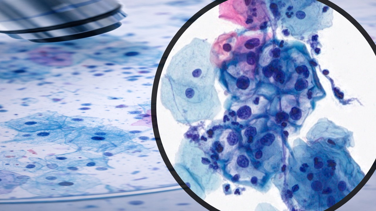
ALPHA PROLIPSIS
LIQUID BASED PAP SMEAR
Our goal is to provide accurate laboratory testing, with a reasonable turnaround time, in a cost-effective manner.
Liquid-based cytology (LBC) is a different way to do the Pap test. Developed in the 1990s, this method presents the following differences compared to the conventional Pap test, resulting in increased sensitivity
Sample collection and transfer
In the conventional Pap test, the cells are collected and smeared directly onto glass slides, and they need to be fixed immediately; usually, using a spray fixative. The problems with this approach are that not all cells are transferred onto the slides, the cells are not in a single layer, the slides can be broken during transportation and the cells can be damaged until they arrive at the lab. In LBC, the cells are placed into a vial with a preservative, which preserves them for weeks and protects them from damage during transport to the lab. The slide, with the cells in a single layer, is prepared in the lab and not by the doctor or nurse.
Sample adequacy
In the conventional Pap test, samples with a lot of blood, mucus and inflammatory cells are typically considered 'unsatisfactory for interpretation' and, as a result, patients are often recalled.
Slide clarity
Conventional slides do not feature a clear background or organize cells in a single layer. These limitations make finding abnormalities harder, which can impact the accuracy of results.
Flexibility
With the conventional Pap test, additional molecular and biomarker tests cannot be run. The ability to run such tests is important in today's environment with evolving guidelines.
Benefits also include:
Health Tips
What is a Pap test?
The Pap test is the most successful laboratory screening method used to identify women who may have a premalignant disease and are at high risk of developing cervical cancer. This is done by collecting cells from the cervix and examining them under a microscope, looking for specific morphologic clues as to the health of the cells present.
Who should have a Pap test and how often?
Recent (2012) guidelines set forth by the American Society for Colposcopy and Cervical Pathology, a consensus document agreed upon by professional and government agencies, recommend the following intervals for screening Pap tests:
No Pap test screening for women aged 20 and younger
Start Pap test screening at age 21
Screen every 2-3 years for women aged 21-29
Co-test with high-risk HPV and Pap test at age 30
If both are negative and patient has not had abnormal Paps or HPV test in last 10 years, extend co-test screening to every 5 years
Can stop routine Pap and HPV co-testing at age 65 if no abnormal history is present
What is the difference between screening and diagnostic Pap tests?
Screening Pap tests are routine tests collected and evaluated for preventive health visits. Diagnostic Pap tests are performed after a patient has been diagnosed with a cervical abnormality and is being actively followed and treated. Diagnostic Pap tests may also be ordered when the patient’s visit is due to abnormal signs or symptoms, such as abnormal bleeding.
How is a Pap test done?
The Pap test is a quick and generally painless procedure. A clinician, usually a doctor or a nurse practitioner, performs this test in the clinic. The clinician inserts a speculum into the vaginal canal. The excess mucus on the cervical opening is removed with a cotton swab, and the clinician gently scrapes a sample of cells from the cervix. The cellular material is either spread (smeared) on a glass slide and fixed, or washed in a liquid-based preparation. The sample of cells is then looked at under the microscope by a board certified cytotechnologist.
Is there anything I should or should not do before my Pap test?
There are a few general recommendations for obtaining optimum results. The test should not be done during menses as menstrual bleeding interferes with the appearance of cells and makes interpretation more difficult. The test should be delayed for 48 hours after the following circumstances:
Sexual intercourse
Use of a douche
Use of a tampon
Use of medicine or spermicidal foams
Use of jellies or vaginal cream
How is a Pap test evaluated?
The Pap test is received in a laboratory from your clinic or doctor. A cytotechnologist then examines the cells using a microscope. If abnormal cells are seen, a cytopathologist (MD) re-examines the abnormal cells and renders an interpretation.
How is a Pap test reported?
The results of the Pap test are reported to the clinician within a week. The clinician would then contact the patient. The Pap test reports follow the standard guidelines outlined by The Bethesda System.
If a Pap test is considered abnormal, does this mean I have cancer?
An abnormal test result does not mean you have cervical cancer, unless it specifically states “Positive for Malignancy.” An abnormal Pap result indicates that there is some degree of cellular change in the cervix’s squamous or glandular cells.
What does this Pap test diagnosis mean?
Negative for Intraepithelial Lesion or Malignancy (NILM): The Pap test revealed no abnormal cell changes. All squamous and glandular cells seen have normal-appearing cellular material. Recommendations will vary depending on your individual risk factors.
Atypical Squamous Cells of Undetermined Significance (ASC-US): Squamous cells do not appear completely normal, but it is uncertain what the cellular changes mean. This diagnosis is suggestive of a squamous intraepithelial lesion but quantitatively and/or qualitatively insufficient for a definitive diagnosis. Sometimes the changes are related to bacterial action, drying artifact or HPV. The recommendation for this diagnosis depends on the patient’s age and clinical history. It will range among repeating in 12 months, reflex HPV testing, or colposcopy.
Atypical Squamous Cells, which cannot exclude a High-grade lesion (ASC-H): Cellular changes of ASC-H do not appear normal, but it is uncertain what the cellular changes mean. Cells categorized as ASC-H have abnormality changes that cannot be definitively diagnosed as high-grade SIL. This diagnosis carries an increased risk of being precancerous. The recommendation for this diagnosis is to perform colposcopy and any other procedures deemed necessary to make a definitive diagnosis.
Low-grade Squamous Intraepithelial Lesion (LSIL): This refers to changes in the size, shape and number of cells on the surface of the cervix. Some of these lesions return to normal on their own without treatment. LSIL is commonly referred to as mild dysplasia or cervical intraepithelial neoplasia 1 (CIN 1). The recommendation varies depending on age, and ranges between repeating the Pap in 12 months or referral to colposcopy and biopsy.
High-grade Squamous Intraepithelial Lesion (HSIL): Cells in this category look very different from normal cells, and they are less likely to return to normal without treatment. They are more likely to develop into cancer if left untreated. HSIL encompasses moderate dysplasia (cervical intraepithelial neoplasia 2– CIN2) and severe dysplasia or carcinoma-in-situ (CIN3). The recommendation for this diagnosis is colposcopy, biopsy, and any other additional procedures to aid in determining the degree of abnormality present.
Advantages of the Liquid Based Cytology
Advantage 1: - Due to the extra sensitivity, there is less likelihood of being recalled for testing.
♦ Advantage 2: - The liquid based test improves the chance of disease being detected and early treatment of pre-cancerous changes on the cervix.
♦ Advantage 3: - The new test reduces the rate of false negative reports when disease is in fact present.
liquid-Based Cytology
An important landmark in the history of the Pap test occurred in 1996 when the U.S. Food and Drug Administration (FDA) approved the ThinPrep™ (Hologic, Marlborough, Mass.) as an alternative to the conventional cervicovaginal smear. This was followed 3 years later by approval of the AutoCyte Prep™ (now known as SurePath™; BD TriPath, Burlington, NC). The newest LBC is the MonoPrep™ (MonoGen, Inc., Lincolnshire, Ill.), which was approved in 2006. LBCs were an important step in the development of automated Pap screening devices—an improved preparation was needed to minimize cell overlap so that automated screeners would perform better in identifying abnormal cells. But LBC performed so well in clinical trials against conventional smears that it found a market independent of auto- mated screening. Although there are exceptions, the great majority of peer-reviewed studies, some of them detailed in this chapter, show an increased detection of low-grade squamous intraepithelial lesions (LSILs) or HSILs with LBC. The debate over increased disease detection with LBC continues, however, and the studies comparing LBC to smears have come under criticism for allegedly sacrificing methodologic purity in their design.26 Nevertheless, LBC offers several clear advantages over conventional smears: the opportunity to pre- pare duplicate slides and even cell block preparations from the residual sample; the option of “out-of-vial” aliquoting for HPV, chlamydia, and gonorrhea testing; an improved substrate for automated screening devices; and a thinner cell preparation that most pathologists and cytotechnologists find less tiring to review than smears.
ThinPrep Pap Test
The practitioner obtains the ThinPrep Pap sample with either a broom-type device or a plastic spatula/endocervical brush combination.
The sampling device is swirled or rinsed in a methanol-based preservative solution (PreservCyt) for transport to the cytology laboratory and then discarded. Red blood cells are lysed by the trans- port medium.
The vials are placed one at a time on the ThinPrep 2000 instrument. The entire procedure (Fig. 1.1A) takes about 70 seconds per slide and results in a thin deposit of cells in a circle 20 mm in diameter (conrast with cytospin: diameter = 6 mm).
A batch-process- ing version (the ThinPrep 3000) is also available. It uses the same consumables (filters and solutions) but allows automated processing of 80 samples at one time. In most cases, only a fraction of the sample is used to prepare the slide used for diagnosis. If needed, the residual sample is available for additional ThinPrep slide preparation, cell block preparation, or molecular diagnostic testing (e.g., high risk HPV, chlamydia, gonorrhea).
A multicenter, split-sample study found that the ThinPrep detected 18% more cases of LSILs and more serious lesions as compared to conventional smears, with no significant difference in the detection of organ- isms. A number of studies have shown significant increases in HSIL detection after the implementation of the ThinPrep.
The ThinPrep is equivalent to the conventional smear in the detection of endocervical AIS.36 Data also show comparable results between ThinPrep slides and conventional smears for the detection of endometrial pathology.
The ThinPrep collection vial has been approved by the FDA for direct testing for HPV, which is particularly useful for managing women whose Pap tests show atypical squamous cells (ASC)
SurePath Pap Test
TriPath Imaging (acquired by Becton Dickinson in 2006) developed the SurePath Pap test (formerly AutoCyte Prep and CytoRich) for samples collected in an ethanol-based transport medium.
In contrast to the ThinPrep and MonoPrep methods, the practitioner snips off the tip of the collection device and includes it in the sample vial.
The equipment to pre- pare slides includes a Hettich centrifuge and a PrepStain robotic sample processer with computer and monitor. The PrepMate™ is an optional accessory that automates mixing the sample and dispensing it onto the density reagent. Red blood cells and some leukocytes are eliminated by density centrifugation. In addition to preparing an evenly distributed deposit of cells in a circle 13 mm in diameter, the method incorporates a final staining step that discretely stains each individual slide.
A multicenter, split-sample clinical trial showed a 7.2% increase in the detection of LSILs and more serious lesions and a significant decrease in the percentage of unsatisfactory specimens.
MonoPrep Pap Test
The practitioner obtains the MonoPrep sample with standard collection devices that are swirled or rinsed in a preservative-filled collection vial, after which the sampling device is discarded. As with the ThinPrep, red blood cells are lysed by the transport medium. The vials are delivered to the laboratory where slides are prepared using the MonoPrep Processor, a fully automated, batch- processing instrument capable of processing 40 samples per hour, with a throughput capacity of 324 samples per 8-hour run. In a split-sample clinical trial similar in design to the ThinPrep and SurePath trials, slides prepared by the MonoPrep method showed a 26% increase in the detection of LSILs and more serious lesions, with no significant difference in relative specificity. MonoPrep provided a significant reduction in unsatisfactory slides, and there was no difference in the presentation of endocervical or transformation zone component or the detection of benign conditions.